PET has the potential to verify charged particle therapy in vivo, reducing uncertainty in particle range and improving treatment accuracy. However, biological washout and the short half-life of positron emitters generated by the treatment beam mean that scans must be performed during or shortly after treatment.
In a new development, a unique transformable PET system that images both during and after irradiation has proved to be feasible. The miniature OpenPET prototype was developed by researchers from the National Institute of Radiological Sciences (NIRS) in Chiba, Japan, led by Taiga Yamaya, PhD.
In "open" mode, the detector ring is sheared along the scanner axis, creating a field-of-view (FOV) centered in the ring that is accessible by the treatment beam. A simple, hand-driven mechanism transforms the detectors into a conventional "closed" toroidal geometry that enables imaging immediately following treatment.
The detector ring comprises 16 detector units arranged in a 250-mm diameter circle. Each unit consists of two depth-of-interaction (DOI) detectors; each of these comprises a 16 x 16 array of Zirconium-doped gadolinium oxyorthosilicate scintillators. In open mode, the detector units are staggered along the scanner axis (Physics in Medicine and Biology, 21 Feb 2006, Vol. 61:4, pp. 1795-1809).
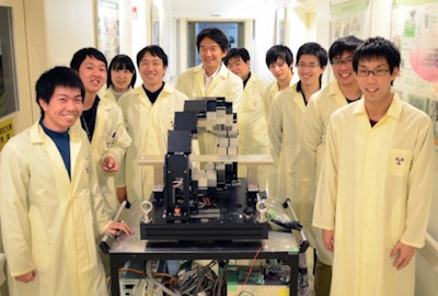 Almost all of the members of the Yamaya laboratory at NIRS.
While conventional PET detectors are attached to a fixed frame, OpenPET detectors are attached to their neighbors by guide rails. Using a rotating handle to drive them along the guides, it takes less than 10 seconds to switch between the two scan geometries. The structure supports a total detector mass of 16 kg. "Designing a transformable mechanism for the heavy detectors was really challenging," Yamaya told medicalphysicsweb.
OpenPET transforms between closed and open scanning modes
Images of a Na-22 point source reconstructed with the 3D-ordered subset expectation maximum (OSEM) algorithm revealed that open mode scans had lower sensitivity and resolution. Sensitivity was 30% lower than in closed mode, dropping from 7.3% to 5.1%. The drop was attributed to a greater detector-phantom distance that decreases the solid angle covered by the detectors.
Quantified using the full-width at half-maximum (FWHM) and the same source, the researchers also observed a lower spatial resolution in open mode (2.6 mm versus 2.1 mm). This difference arose from different detector orientations relative to the center of the field-of-view (FOV). In closed mode, the scintillators point toward it, while in open mode, they point 45° away from the center of the FOV, introducing a parallax error. Based on the results, the researchers propose maximizing OpenPET's performance by imaging patients in open mode during treatment, then in closed mode immediately following treatment.
In another experiment, the team delivered 2.5 Gy at the Bragg peak to a cuboid plastic phantom using a carbon-11 beam from NIRS' HIMAC (Heavy Ion Medical Accelerator in Chiba). This revealed that sufficient count statistics for image reconstruction were achievable upon combining counts acquired during irradiation with less than a minute of acquisition following irradiation. In this way, the system improves upon previous OpenPET designs that could only be operated in open mode.
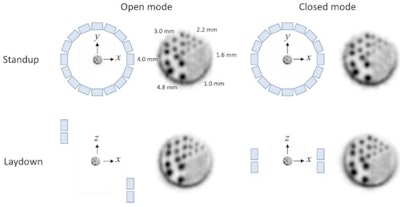 One of two experiments measuring OpenPET's spatial resolution using a rod phantom standing up (top) and laying down (bottom). Using this test, there was no difference between scanning modes; both were used to resolve rods 2.2 mm apart.
"It is hard to satisfy all imaging requirements at the same time with one mode," said first author Hideaki Tashima, PhD. "Therefore, the transform system works well -- we can minimize the sensitivity loss in practical use by switching between the two modes." The dual-mode scanner also saves space and is more economical than two separate PET systems.
The in-beam experiment showed that the prototype could successfully resolve a shift in Bragg peak depth when half the beam was fired through a 9-mm thick PMMA board. It also demonstrated agreement between the measured position of the Bragg peak and that predicted by the planning system within 2 mm, the uncertainty of the experiment.
Based on their findings, the researchers are developing a larger clinical system and plan to acquire the first patient images later this year. A major challenge faced by the team is design of a robust structure that allows movement of 80 kg of detectors between scanning modes.
© IOP Publishing Limited. Republished with permission from medicalphysicsweb, a community website covering fundamental research and emerging technologies in medical imaging and radiation therapy.


