It's more than 500 years since the birth of probably the most famous and influential anatomist who ever lived. Andreas Vesalius was born in 1514 and studied medicine in Paris, and at the University of Louvain in Belgium as well as in Padua, Italy. Immediately upon graduation from Padua, he was appointed to the chair of anatomy and surgery at the age of 23.
His early interests were in venesection (blood-letting) and he contributed early literature on this subject. He was a pioneer experimenter and performed many dissections and encouraged his students to do so to help with understanding the structure of the human body. He challenged many of Galen's findings (for many years no one had dared challenge the prevailing orthodoxy). Vesalius claimed Galen had not dissected humans and that his findings had been based on animal anatomy.
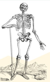
Vesalius was an excellent teacher and initially created anatomical drawings for his students on woodcut posters. Woodcut and copperplate illustrations were popular in this era and were used to visually demonstrate knowledge about the cosmos and the new cartographic discoveries about the planet that explorers were making at the time. Examples included Frisius' Cosmographia in 1529, which replaced the outdated Ptolemic maps, and Leonard Fuchs' botanical masterpiece from 1542 De historia sterpium.
Vesalius's meticulous dissection and acute observational powers enabled him to make many new discoveries that disproved Galen's teachings. He showed the lower jaw had only one bone, the sternum had three parts, and the sacrum several segments amongst many new anatomical discoveries.
Vesalius also showed that there was no communication between the ventricles of the heart as Galen had thought. He showed the heart had four chambers: two atria and two ventricles and described the azygous vein. He made major discoveries in the vascular and circulatory system that challenged previously held beliefs.
Vesalius is probably best remembered today for his monumental anatomical work De humanis corporis fabrica published in 1543 with illustrations possibly done by artists from Titian's school. The title page depicts a crowded anatomical theater with everyone watching the dissection in the center. Galen, Hippocrates, and Aristotle are symbolically represented as the giants of the time, but men who had not corroborated their findings with direct observation. Vesalius was eventually to join the pantheon of revolutionary medical men.
Since its publication, anatomical errors have been unearthed in the masterwork -- notably in the anatomy of the eye with inaccurate illustrations of the eye muscles, probably because he had dissected an animal eye rather than a human one.
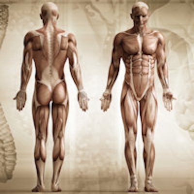
Nevertheless, the book was, and still, remains an extraordinary achievement and one of the finest anatomical/medical books ever written. The illustrations of the dissected bodies are works of art in their own right. The depiction of human musculature is extraordinary in their accuracy and detail and has never been bettered. The displayed structures are profusely annotated.
The book opened up new vistas of knowledge about the human body structure as never seen before. Anatomy subsequently became a legitimate subject of study for future artists as well as doctors so this book has inspired generations of artists as well as doctors. Ironically, following the publication of his magnum opus Vesalius resigned his chair in Padua and became physician to Charles V, ruler of the Roman Empire (1519-1556) and to whom he dedicated his book. Vesalius died following a shipwreck off the Greek islands in 1564, only 49 years old.
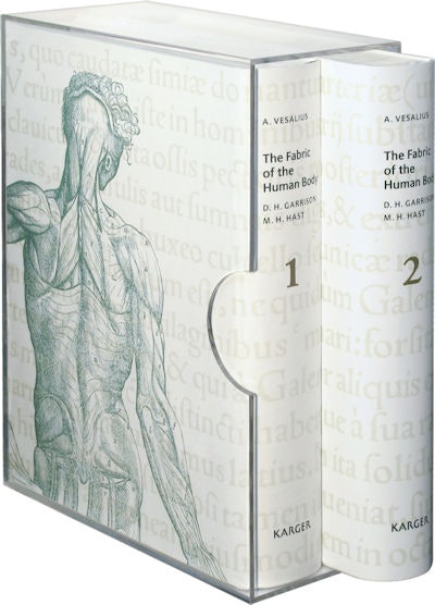 The Fabric of the Human Body was published by Karger in 2014.
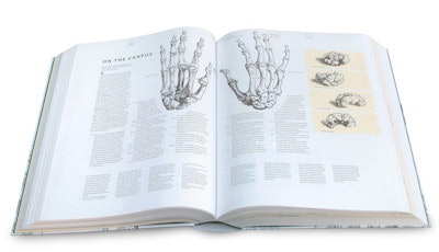 Inside the book.
Radiologists, whose work relies on knowledge of living anatomy, would find the book of great interest. Although it is not used today by current doctors for learning anatomy, the illustrations remain remarkable. Today, we are able to look inside the body without dissecting, and much anatomical teaching in medical schools is now performed using radiological imaging and reconstructions that are easily performed using modern CT scanners and MRI machines.
It is indeed remarkable how today images of the body's musculature and vascular system can be produced without a knife touching the skin. Many consider these current radiological images of the human body from these machines as works of art in their own right as exhibitions by Franz Fellner, an Austrian radiologist, will testify.
Last year saw the publication by Karger of the beautifully annotated translation of his masterpiece The Fabric of the Human Body so modern readers can gain insight into probably the most famous medical book ever published. The 1,400-page book is in two volumes and is an annotated translated version of the 1543 and 1555 editions. It is indeed a scholarly fitting tribute to Vesalius and his pioneering magnum opus.
Dr. Arpan K. Banerjee, MBBS (LOND) FRCP FRCR FBIR is chairman of the British Society for the History of Radiology and co-author with Dr. Adrian Thomas of The History of Radiology.
References
- Andreas Vesalius Wikipedia page. Wikipedia website. http://en.wikipedia.org/wiki/Andreas_Vesalius. Accessed 24 December 2014.
- Garrison DH. Vesalius and the achievement of the Fabrica. Karger Gazette. 2013: 73, 2-4.
- Vesalius A. Fabric of the Human Body. Basel, Switzerland: Karger; 2014.
All images courtesy of Karger.
The comments and observations expressed herein do not necessarily reflect the opinions of AuntMinnieEurope.com, nor should they be construed as an endorsement or admonishment of any particular vendor, analyst, industry consultant, or consulting group.


