The history and archaeology of ancient Egypt has fascinated us for many years. The interest was stimulated by Napoleon Bonaparte's campaign in Egypt and Syria from 1798 to 1801. There was a scientific aspect to this military expedition and Napoleon took along with him 167 scholars and scientists. Part of his aim was to spread the principles of the Enlightenment.
Many discoveries were made including those of the young engineering officer, Pierre-François-Xavier Bouchard, who discovered the Rosetta Stone in July 1799. It was the Rosetta Stone that allowed us to understand ancient Egyptian hieroglyphics.
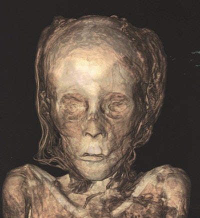 CT scan showing the remarkable preservation of the facial features. The eyelids, lips, and nose are preserved, and the hair is clearly seen. All colored illustrations from Taylor JH, Antoine D. Ancient Lives New Discoveries: Eight Mummies, Eight Stories. London: British Museum Press; 2014.
This developed into the Egyptomania of the 19th century, with a fascination for all things Egyptian. Many mummies were unwrapped and much information was obtained. However, in unwrapping, the mummy is basically broken up. It is fortunate that the collection of mummies in the British Museum remained intact.
It was therefore obvious that when x-rays were discovered, that some of the earliest items radiographed were these ancient mummies. For example, on 23 October 1896, Charles Thurstan Holland radiographed a mummy bird. This took a three-minute exposure and he said that it was "a relief to have something to examine that would keep still and which was not frightened by our apparatus, the sparks, and so on."
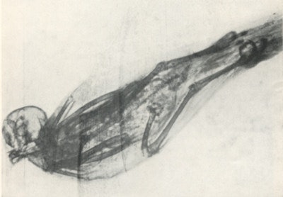 Mummy bird from an Egyptian tomb, taken by Charles Thurstan Holland on 22 October 1896.
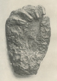
William Meadowcroft in his The ABC of the X-rays from October 1896 illustrates a photograph of a mummified hand of an Egyptian princess, which had been obtained from the tombs of the Kings in Thebes in 1892. The radiograph shows excellent bony detail. The contents of the mummy can therefore be examined without destroying it in the process. In those early days, the apparatus was of low power, however small items could be examined. Many of the early x-ray books have illustrations of the examination of this type of material.
As radiology advanced so these newer techniques were applied to Egyptology. This could include radiography of an entire human mummy, which took place in the 1960s and 1970s as good portable apparatus became available. From the 1990s, many mummies were examined with CT, and increasingly detailed images have been obtained. This means that the need to unwrap the mummy is avoided.
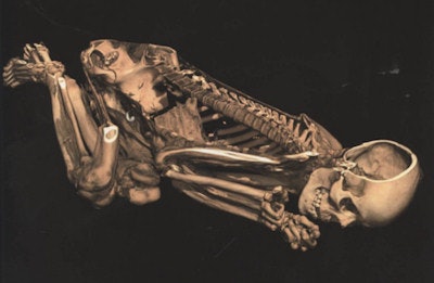
There is an exciting exhibition at the British Museum titled Ancient lives new discoveries: Eight mummies, eight stories. These eight mummies are examined using modern scanners and produce astonishing images. The older x-ray techniques have been replaced by the latest generation of dual-energy CT scanners. In a similar way to the older physical unwrapping, the modern CT scan can produce a virtual unwrapping, removing the various layers of the body down to the skeleton.
This reveals detailed biological information. The information was analyzed using methods developed by forensic archaeologists and physical anthropologists. Information about diet, the state of health, and the embalming techniques that were used are obtained. In the exhibition, the eight mummies are displayed with supporting material including the CT scan. The CT images are projected and can be virtually unwrapped and manipulated by the visitor using a touch-sensitive controller.
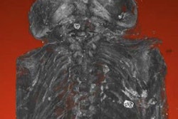
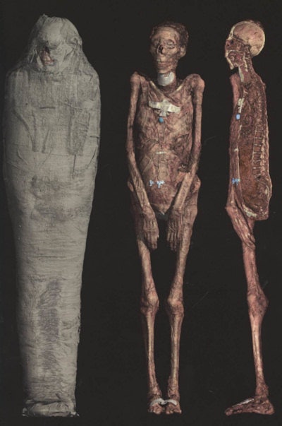 Modern CT scan showing the virtual removal of the wrappings illustrating the mummified remains. The sectioned image on the right shows the internal structures.
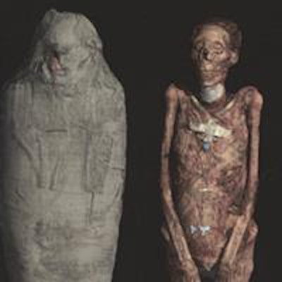
The CT can be directly compared with the adjacent mummy. I found the experience most remarkable. It emphasized to me the essential humanity of the mummies that were being examined, with the images looking like the CT scans that are reported every day. We are therefore looking at human beings who are the same as we are, and we are not looking at museum objects. These are people who lived and breathed and had feelings in the same manner that we do.
In the collection, two of the mummies are naturally mummified in the warm dry atmosphere of Egypt, and the other six are artificially embalmed. Considerable information can be obtained from the scans with information about age, lifestyle, and any diseases. In one of the mummies, calcific atheroma is clearly visibly as is poor dentition.
 Sectioned image of a naturally mummified body.
One of the advantages of radiology is that the internal contents of the mummy can be precisely determined. For example, on the outside, a bird mummy might contain a fish, and an apparently female mummy can be shown to be male.
I came away with the feeling that we have only started to scratch at the surface of the knowledge that we can gain using radiology.
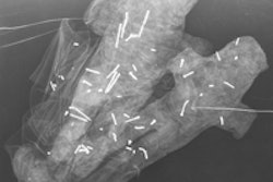
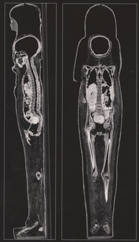 Longitudinal CT sections of mummy in its cartonnage case.
The exhibition is at the British Museum in London and finishes on 30 November 2014. It is highly recommended and anyone interested in Egyptology or modern scanning will be as fascinated as I was.
Dr. Adrian Thomas is chairman of the International Society for the History of Radiology and honorary librarian at the British Institute of Radiology.
Reference
Taylor JH, Antoine D. Ancient Lives New Discoveries: Eight Mummies, Eight Stories. London: British Museum Press; 2014.
The comments and observations expressed herein do not necessarily reflect the opinions of AuntMinnieEurope.com, nor should they be construed as an endorsement or admonishment of any particular vendor, analyst, industry consultant, or consulting group.


