Elaitra, the U.K./U.S. company specializing in accelerated reading for digital breast tomosynthesis (DBT), has released an AI-enabled product that aims to dynamically map the same tissue site between views.
The firm will highlight its ViewFinder product at RSNA 2025 from 30 November to 4 December in Chicago and at the San Antonio Breast Cancer Symposium (SABCS) from 9 to 12 December in Texas.
ViewFinder, which has been 510(k)-cleared by the U.S. Food and Drug Administration (FDA), offers dynamic matching to assist in correlating findings across views -- mapping the same tissue location between DBT views and slice levels, with a confidence-scaled ellipse highlighting the corresponding location. It also provides reference measurements and coronal views.
The AI-enabled tool is vendor-agnostic and can be deployed on-premises or through approved cloud providers. It can also be paired with low-latency cloud DBT reading where appropriate, Elaitra added.
EJR study
Results from a retrospective, two-part feasibility study led by Stephen Morrell, PhD, Elaitra CEO and former AI research scientist at King’s College London, have been published in the November edition of the European Journal of Radiology. The findings showed that the use of the tool “significantly improves spatial localization accuracy in DBT, particularly for nonexpert radiologists interpreting complex cases.”
The first part of the study was a subjective evaluation by 14 radiologists of the tool’s usefulness and impact on their confidence; in part two, 12 radiologists marked lesion annotations with and without assistance from the tool following a washout period.
The researchers noted that examination without the AI-based tool’s assistance resulted in 32% larger mean localization errors in abnormal cases (p = 0.049), with 37.5% larger maximum distance errors (MDE) (p = 0.036). They observed the most notable improvements occurring in the most difficult cases (defined as above 90th percentile), with MDEs dropping from 48.1 mm to 26.2 mm for nonexpert readers.
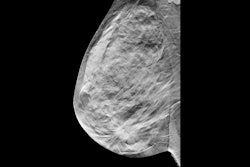
 Example case demonstrating improved accuracy in lesion location with ViewFinder (VF) on versus VF off. The mediolateral oblique (MLO) view shows the source reference-standard annotation (red), while the craniocaudal (CC) images show individual radiologist annotations (blue) for both, ViewFinder ON and OFF, and the reference standard annotation
(in red). Courtesy of Stephen Morrell, PhD, et al and EJR. (Note: For interpretation of the references to color in this figure legend, the reader is referred to the original version of this article.)
The gains for nonexpert readers from the interactive system that “can meaningfully reduce spatial errors” are particularly important, the researchers add, because nonexpert radiologists interpret up to 70% of mammograms in the U.S.
As most screen-detected lesions are small, and DBT does not inherently solve cross-view mental matching, the ViewFinder’s ability to bring the match within lesion bounds is significant for diagnostic targeting, increasing the diagnostic accuracy of workups, and improving communication, Elaitra added.
The vendor is currently recruiting pilot sites to further validate outcomes. Also, it is recruiting radiologists for an international follow-up study aimed at expanding generalizability and outcomes data.
The EJR study is online here.