In groundbreaking research, a group from Bordeaux, France, has used a basic MRI method to measure iron content in the substantia nigra to obtain a better reading on how stroke affects the brain and long-term patient outcomes. The results were published online on March 12 in Radiology.
Called R2* mapping, the MRI technique can detect increased iron concentrations in the substantia nigra, and it can be added to follow-up MRI scans to monitor neurodegeneration and resulting poor motor function among stroke patients.
"Although secondary neurodegeneration of the substantia nigra has been previously proposed, no insight on the potential clinical relevance of this phenomenon has previously been reported," wrote lead author Dr. Pierre Antoine Linck and colleagues from Centre Hospitalier Universitaire in Bordeaux. "Taking advantage of quantitative R2* mapping, we provide preliminary evidence for a relationship between substantia nigra R2* changes and long-term outcome after stroke."
Brain connections
The substantia nigra is associated with motor control, emotions, cognition, and motivation. Given its connections to the supratentorial region and other parts of the brain, it is thought that infarctions occurring in other areas can adversely affect the substantia nigra. However, whether these nearby or distant infarcts cause degeneration of the substantia nigra after a supratentorial stroke has not been studied previously.
Iron is a known contributor to normal brain function, but too much iron can lead to neurodegeneration. Coincidentally, MRI is particularly helpful in detecting changes in iron concentration in the brain through R2* mapping.
Until the current study, however, determining degeneration in the substantia nigra could only be done through postmortem brain exams. Thus, as the researchers wrote, "Better techniques are required to understand the impact of supratentorial injury on the substantia nigra after stroke."
To test the efficacy of R2* mapping relative to assessing iron content and neurodegeneration, Linck and colleagues consecutively enrolled 181 patients between June 2012 and February 2015. The participant group included 127 men and 54 women with a mean age of 64.2 years (± 13.1 years) who presented with suspected minor to severe supratentorial cerebral infarcts (75 striatum infarcts and 106 in other brain locations). The subjects were evaluated with diffusion-weighted MR imaging 24 to 72 hours after their event.
MRI scans were performed on a 3-tesla scanner (Discovery MR 750w, GE Healthcare) with a 32-channel phased-array head coil. The researchers used the same protocol for baseline and one-year follow-up imaging, which included diffusion-weighted imaging (DWI) and corresponding apparent diffusion coefficient values, 3D T1-weighted images, and 2D T2* multiecho fast gradient echo sequences. R2* maps were computed from multiecho T2*, followed by coregistration of all sequence maps to the native 3D T1 images.
The value of R2*
At the one-year follow-up, the researchers found higher average R2* values within the substantia nigra after a supratentorial infarction, "a finding that we interpret as iron accumulation as a result of neurodegeneration," they wrote. In addition, R2* maps consistently showed higher substantia nigra R2* values (iron concentration) when the infarct occurred at the ipsilateral striatum compared with R2* values when the infarct occurred in other locations. In other words, R2* mapping of iron content also could be used to image neurodegeneration remotely, if strokes were to occur in disconnected areas.
"This finding could be clinically useful because it shows that a simple magnetic resonance imaging method such as R2* can provide a more comprehensive picture of the consequences of an infarct," added Dr. Thomas Tourdias, PhD, study co-author and professor of radiology, in a statement.
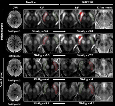 MR images show examples of visual R2* modifications within substantia nigra at baseline (24-72 hours after stroke) and at one-year follow-up in striatum (participants 1 and 2) and control groups (participants 3 and 4). Axial R2* maps are enlarged at mesencephalon level and one of the individual T2* echoes also is shown at follow-up. The substantia nigra is segmented in red ipsilateral to infarct and in green contralateral to infarct. Images without outlines are also shown. Brighter R2* spots are noted with red arrows. Asymmetry index of 95th percentile of R2* within substantia nigra (SN-AI95) measured in these participants is indicated for reference. Participants 1 and 2 show marked asymmetry of R2* within ipsilateral substantia nigra at follow-up, compared with baseline after infarct involving entire striatum (participant 1) or limited to putamen (participant 2). Asymmetry also is visible on individual T2* echo at follow-up. Conversely, no asymmetry was observed in participants 3 and 4 sparing striatum. Images courtesy of Radiology.
Linck and colleagues also observed an association between high iron content and poor long-term physical outcomes for stroke patients, particularly when increased iron levels were found on the same side of the brain where the infarction occurred. For example, follow-up tests showed reduced performances of patients when they tried to use their nondominant arms and in a 10-mile walking test.
"To our knowledge, for the first time, we demonstrated that degeneration of the substantia nigra may be associated with less favorable clinical outcome," the authors noted.
Given the results, the group suggested that R2* mapping could be added to follow-up MRI scans after a stroke to monitor neurodegeneration by assessing the iron levels in the substantia nigra. In more general terms, the findings support the use of iron imaging as a marker for neurodegeneration, which also currently is being explored for neurodegenerative diseases such as Parkinson's.

