Below is a list of the finalists for the second edition of the EuroMinnies, AuntMinnieEurope.com's annual awards scheme.
The finalists were drawn from a total of 79 semifinal candidates in eight categories, based on nominations submitted in late 2019 by members of AuntMinnieEurope.com. View the full list of semifinalists.
One winner will be selected for each category by our expert panel. The winners will be announced in February, and the trophy presentations will take place at ECR 2020 in Vienna.
Most Influential Radiology Researcher
Prof. Christiane Kuhl, PhD, RWTH Aachen University Hospital, Aachen, Germany
Meeting organizers often tend to hold back their star presenter until the end of a conference, so it was no surprise that Prof. Christiane Kuhl, PhD, gave the closing lecture at the 2019 International Society for Magnetic Resonance in Medicine (ISMRM) annual meeting in Montreal. She made the most of the opportunity, delivering a typically lucid and vigorous call to action.
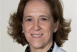
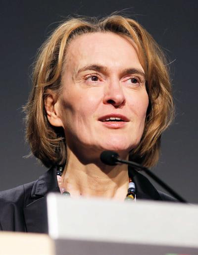 Prof. Christiane Kuhl, PhD.
Her core message was that better risk assessment methods and more appropriate use of techniques such as MRI open the door for personalized cancer screening strategies. The approach is to adapt the screening efforts closely to an individual woman's lifetime risk of developing breast cancer, thereby improving the success of screening programs.
"We now have the predictive measures, and we as radiologists will contribute to improved risk prediction through imaging biomarkers that we provide," noted Kuhl, who was voted runner-up in the Most Influential Radiology Researcher category for the 2019 EuroMinnies. "And we have the tools to meet this need. Let us begin to tailor our preventive efforts accordingly."
Underdiagnosis is the real issue in breast cancer, not overdiagnosis, because the adverse effects of overdetection can be avoided by better patient information and management. The bigger problem is underdiagnosis -- not detecting the aggressive cancers that should be found. Better selection criteria for breast cancer screening programs beyond just sex and age are vital to future success, and abbreviated MRI can expand the use of breast MRI, she said in her ISMRM lecture.
Other accolades have followed. On 30 September 2019, Aachen University Hospital announced that Kuhl had been accepted into the Leopoldina, the National Academy of Sciences, due to her achievements in imaging and image-guided tumor treatment.
"I am very much looking forward to the professional exchange, but above all to the opportunity to discuss the social role of researchers and research," she said. "In a polarized public sphere that reduces things to simple truths, scientists are increasingly perceived as foreign objects that have fallen out of time. Helping to change that -- I consider this an important task!"
The European Society of Breast Imaging (EUSOBI) awarded its Gold Medal to Kuhl in late 2018. Also, she received the Impact Award from the U.S. National Consortium of Breast Centers, an interdisciplinary specialist society.
Prof. Mathias Prokop, PhD, Radboud University, Nijmegen, the Netherlands
When you think of prominent European radiologists involved in lung cancer screening, the chances are high that Prof. Mathias Prokop, PhD, will come to mind. He is widely known as a leading advocate of low-dose CT screening for lung cancer in high-risk patients.
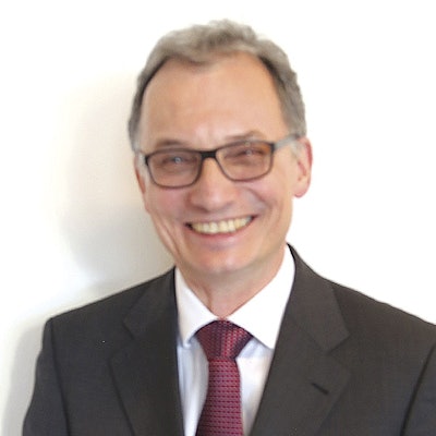 Prof. Mathias Prokop, PhD.
For more than two decades, Prokop has conducted cutting-edge research in chest screening with CT. As early as 2007, he was co-author of an important article in Radiology titled "Monitoring of smoking-induced emphysema with CT in a lung cancer screening setting: Detection of real increase in extent of emphysema." He has written more than 250 peer-reviewed articles, 50 book chapters, 300 scientific abstracts, and 300 invited lectures, and he has published a textbook on spiral and multislice CT, according to his bio on the European Society of Radiology (ESR) website.
At ECR 2017, Prokop gave the Josef Lissner Honorary Lecture: "The future of CT: From hardware to software." Another career highlight was his close involvement in the Dutch-Belgian NELSON (Nederlands-Leuvens Longkanker Screenings Onderzoek) study, which showed that mortality decreased by 26% among men and by 61% among women after CT lung screening.
"Lung cancer screening appears to work better among female patients. This is probably because comorbidity among women is lower, and they more frequently have slow-growing tumors," he told delegates at the 2019 Bayerisch-Österreichischer Röntgenkongress in the influential Holzknecht Lecture.
"If we screen for long enough, the optimum effect on survival will develop," said Prokop, who is head of radiology and nuclear medicine at Radboud University Nijmegen and professor of radiology at UMC Utrecht. "It just takes a while until all tumors within the population have been captured and for new tumors to have been detected early enough."
Life-extending treatment procedures for advanced tumor stages, such as immunotherapy, work well, but because they are expensive, the cost per life saved by a screening program is high compared to CT. "The only chance of a cure is to diagnose lung cancer as early as possible," he noted. "Can we afford not to offer screening?"
In a video interview with AuntMinnieEurope.com at ECR 2019, he explained that finding the right partners is crucial in research.
"If you want to have a significant impact, you have to do it together with others, and only then will you succeed," he said. "Having a good idea is not really enough for success in research -- you have to make sure it has a potential impact too."
Most Effective Radiology Educator
Dr. Erik Ranschaert, PhD, European Society of Medical Imaging Informatics (EuSoMII) & ETZ Hospital, Tilburg, the Netherlands
Hype and technical jargon are all too common in the world of medical computing, but Dr. Erik Ranschaert, PhD, has a rare ability to address complex issues and describe difficult concepts in a clear and understandable way. Like all good educators, he can convey a message with skill and simplicity -- whether it's in the form of an oral lecture, written article, video interview, or podcast.
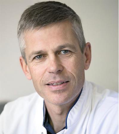 Dr. Erik Ranschaert, PhD.
Those with long memories will recall Ranschaert's popular magazine column, "Surfing the Net," in the late 1990s. Each month he described several websites that had caught his attention, and his observations and predictions were extremely astute. For example, soon after Google.com first arrived on the scene, he identified the site as a valuable search engine that could allow users to navigate the internet.
He became chairman of the ESR Computer Applications Subcommittee in 2008 and was a member of the ESR eHealth and Informatics Subcommittee between 2014 and 2016. He spoke and wrote with authority and insight on PACS and informatics, dealing with hot topics such as data security and the use of mobile devices and social media.
But it's since the evolution of artificial intelligence (AI) that Ranschaert's skills as a communicator and organizer have become apparent. As vice president of EuSoMII between 2016 and 2018, he helped run conferences and seminars about AI. He was elected president of the society in November 2018, and he played a key role last year in the issuing of a global statement calling for codes of ethics and practice governing the use of AI.
In October 2019, Ranschaert helped EuSoMII reach an agreement with the Society for Imaging Informatics in Medicine (SIIM) under which the societies pledged to collaborate more closely and to promote imaging informatics around the world.
"It is an absolute honor and pleasure to make this great step forward in strengthening the position and importance of imaging informatics in medicine, especially for the younger generation," he said. "We have to join forces in order to improve digital innovation for the benefit of the patient."
Ranschaert still thinks there's an urgent need for radiologists to become better informed about AI, as he outlined in a video interview at ECR 2019. Along with Prof. Sergey Morozov and Dr. Paul Algra, he has edited a book called Artificial Intelligence in Medical Imaging: Opportunities, Applications and Risks.
Prof. Laura Oleaga Zufiría, PhD, European Diploma in Radiology & Hospital Clinic of Barcelona, Barcelona, Spain
Prof. Laura Oleaga Zufiría, PhD, is scientific director of the European Diploma in Radiology (EDiR), as well as being immediate past chair of the ESR Education Committee and a past member of the ESR Executive Council. Since 2009, she has worked as head of radiology at the Hospital Clinic of Barcelona. Her own area of special interest is MRI, especially in head and neck imaging and neuroradiology, but improving education across Europe is her raison d'être.
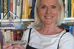
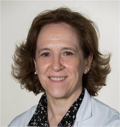 Prof. Laura Oleaga Zufiría, PhD.
"Training and education are aspects of my profession that have always interested me. Since I began my residency in radiology I have been interested in training and I have always worked and enjoyed working with students and residents," Oleaga Zufiría explained in an interview on the ESR website in 2015. "My major motivation is the utmost importance of investing in education if we want to progress in the profession, and continued education in radiology is an essential aspect in this regard."
Under her leadership from 2015 to 2018, the ESR Education Committee did much to promote training in radiology and to achieve homogeneity, excellence, and quality. Its stature has grown as a consultative body for all educational activities within the ESR, working closely with the European School of Radiology (ESOR), the ESR e-learning platform, and the European Board of Radiology (EBR), which organizes the EDiR.
"The participation of trainees in the development of educational content such as the e-learning platform is essential as they represent the most important target group of the educational activities organized by the ESR," Oleaga Zufiría commented.
At EDiR she has coordinated the development of several tools to prepare for the diploma, including EDiR - The Essential Guide. This book was created and reviewed directly by a team of experts from the EBR, and she wrote it along with Drs. Judith Babar, Oguz Dicle, Hildo J. Lamb, and Fermín Sáez.
Serving patients and doctors better, along with meeting their expectations, is one of her main goals, and she emphasizes the need to promote technological research to achieve better healthcare. To establish good working relationships, it is necessary to be direct and to promote collaboration among all professionals involved in patient care, and humility and honesty are essential in our profession, she believes. In addition, she has a personal interest in languages and linguistics.
In August 2017, Oleaga Zufiría was onsite at the Hospital Clinic to take control of the imaging effort following a terrorist attack in Barcelona. Fourteen people were killed and more than 130 were injured after a van drove into pedestrians. Within an hour of the attack, four interventional radiologists performed lifesaving embolization procedures on three patients with severe abdominal hemorrhages.
Radiology Rising Star
Dr. Luísa Costa Andrade, Champalimaud Foundation, Lisbon, Portugal
Since May 2019, Dr. Luísa Costa Andrade has worked as a gastrointestinal radiologist and a member of the Pancreas Fast Track program (multidisciplinary team) at the Champalimaud Foundation, a private biomedical research foundation created in 2004 after the death of entrepreneur António de Sommer Champalimaud. It undertakes research in the fields of neuroscience and oncology, and the Champalimaud Clinical Centre provides specialized treatment for cancer patients.
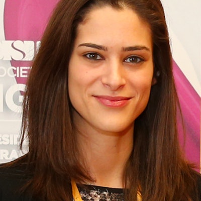 Dr. Luísa Costa Andrade.
Costa Andrade completed her radiology training in 2016 at the Hospital and University Center of Coimbra in 2016, after which she had spells at the Hospital Particular Alvor (HPA group) in Alvor, Portimão, and at the University Hospital Centre of the Algarve, Portimão/Faro. She was secretary and board member of the ESR Radiology Trainees Forum (RTF) from March 2015 to March 2017.
"The main achievement was the approval of the terms of reference by the executive board of the ESR, which gave the RTF the status of an ESR subcommittee," she said in an interview with the ESR in 2017. "Other achievements were the direct representation of the RTF on various ESR committees and subcommittees, the successful conduct of several surveys with a high number of participants, collaboration with the ESR Education Committee in the review of the internship and undergraduate curricula, and improving and promoting the e-learning platform as well as the Invest in the Youth and Rising Stars programs."
Costa Andrade was president of the National Portuguese Committee of Radiology Residents from 2014 to 2016 and a member of the Portuguese Committee of Radiology Residents from 2012 to 2016. She has been a member of the abdominal section of the Portuguese Radiological Society since May 2017.
She has worked overseas too. In 2015, she completed a two-month fellowship on abdominal MRI at the Academisch Ziekenhuis Maastricht (azM) in Maastricht, the Netherlands. Costa Andrade specialized in research on diffusion-weighted MRI of rectal cancer and colorectal-liver metastases. Also in 2015, she did a three-month fellowship on abdominal and digestive MRI at Hôpital Erasme, Université Libre de Bruxelles, Brussels.
Her mentors have included Prof. Filipe Caseiro Alves from Coimbra, Prof. Regina Beets-Tan from Amsterdam, and Prof. Celso Matos from Lisbon, and she said she has been inspired by the ECR, particularly the European Excellence in Education program and the Rising Stars program for residents and students.
"The Invest in the Youth program allows several interns to have the opportunity to make their work known and to be present at one of the largest radiology congresses in the world and assumes itself as one of the ECR's greatest assets," she added.
Dr. Tsvetelina Teneva, Medical University of Varna, Varna, Bulgaria
Dr. Tsvetelina Teneva is an assistant professor of radiology in the department of diagnostic imaging and radiotherapy at the Medical University of Varna in Bulgaria. The department is headed by Prof. Boyan Balev, PhD.
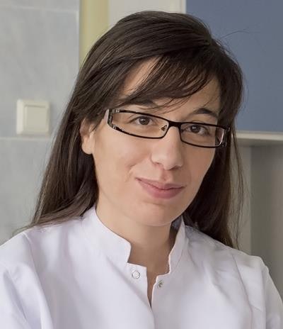 Dr. Tsvetelina Teneva.
She is a past member of the ESR Radiology Trainees Forum. She was a speaker at Radiology Together, the 6th Meeting on Emergency Radiology, held at the Acibadem City Clinic Tokuda Hospital in Sofia in June 2017.
At ECR 2018, Teneva was co-author of a digital poster put together by a team led by Dr. Ralitsa Popova from the Medical University of Varna. Called "Imaging features of extramedullary hematopoiesis (EMH)," it focused on the pathophysiology of the process and how to identify the typical and less common locations for proliferation of hematopoietic cells outside the bone marrow. It also presented findings on x-ray, CT, and MRI indicative of EMH and discussed the specific features that distinguish EMH from other pathologies.
"Extramedullary hematopoiesis refers to the proliferation of hematopoietic cells in organs and tissues other than the bone marrow. Blood develops initially within the core of 'blood islands' in the mesoderm, both within the embryo and outside the embryo (extraembryonic mesoderm). During the fetal development, the hematopoietic stem cells are located in different tissues (e.g., liver, spleen, thymus, etc.). In the adult the stem cells are located in the bone marrow," the authors noted.
At ECR 2017, Teneva was co-author of another poster, "Viral encephalitis: A review," for which the lead author was Dr. Emilian Kalchev.
"Neuroradiological imaging techniques, especially MRI, may contribute important information for distinction between brain infection and others (e.g., other causes of brain inflammation or ischemia), but they contribute only little to the determination of the type of virus," the team wrote. "However, there are some encephalitic viral infections with neuroradiological specifics which may help to isolate the differential diagnosis."
Most Significant News Event in European Radiology
European societies join in ethical statement on artificial intelligence
Over the past couple of years, the medical imaging community has realized that it needs to consider the potential impact of automation more deeply and work out an ethical approach to artificial intelligence. Get your ethics right and machines will not take over and dictate matters -- at least that's how the theory goes. But what exactly does this mean in practice?

In 2019, seven European and U.S. societies came together to produce a new statement calling for codes of ethics and practice governing the use of AI. They agreed that the ethical use of AI in radiology must promote well-being, minimize harm, and ensure the benefits and harms are distributed justly among the possible stakeholders. The overall aim is to promote any use of AI that helps patients and block the use of radiology data and algorithms for financial gain.
"We believe AI should respect human rights and freedoms, including dignity and privacy," the authors wrote in the final statement. "It should be designed for maximum transparency and dependability. Ultimate responsibility and accountability for AI remains with its human designers and operators for the foreseeable future."
The document contains details about specific ethical considerations related to data, algorithms, and practice. Establishing these regulations, standards, and codes of conduct to produce ethical AI means balancing the issues with appropriate moral concern, and achieving ethical AI will require all parties to gain trust in the technology.
"To ensure the safety of patients and their data, AI tools in radiology need to be properly vetted by legitimately chosen regulatory boards before they are put into use," the authors noted. "This requires both radiology-centric AI expertise and technology to verify and validate AI products."
They also emphasize the need to continuously update and monitor the regulations and codes of conduct and regularly reassess this topic. They realize an important start has been made, but the job is not yet complete.
Health bosses say shortage of radiologists puts lives at risk in U.K.
Coping with insufficient numbers of radiologists is a difficulty faced in many European countries, but the situation has become particularly acute in the U.K. over recent years. In April 2019, the publication of new figures underlined the scale of the problem and how staff shortages were posing a serious danger to patient safety.
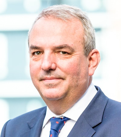 Prof. Mark Callaway.
"Service leaders are now telling us loud and clear that staff shortages are putting patients at risk, with three-quarters saying they cannot guarantee a safe service," noted Prof. Mark Callaway, lead author of the annual radiology workforce report of the Royal College of Radiologists (RCR).
"Health boards desperately need to recruit, but our report shows that new consultant numbers and overseas and locum appointments are just not enough to plug staffing gaps. As a result, our radiologists are exhausted and the NHS [National Health Service] bill for outsourcing scan reporting continues to climb," he added.
The RCR collected data and comments from imaging managers from the U.K.'s 172 health boards and NHS trusts/hospital groups that employ radiologists. It found the following:
- NHS hospitals spent 165 million pounds (196 million euros) in 2018 on outsourcing, overtime, and locums to cover radiologist scan reporting -- 49 million pounds (58 million euros) more than in 2017 and three times what was spent in 2014.
- The amount spent on outsourcing would pay for 1,887 full-time radiologists, which would more than pay to cover the current shortfall of 1,104 consultants.
- Only one in five trusts and health boards has enough interventional radiologists to run a safe 24/7 service to perform urgent procedures.
The report shows the number of NHS radiologists is falling well short of demand, and respondents confirmed that staff shortages are leading to delayed cancer diagnoses and inadequate emergency diagnostic and interventional services.
The situation does not seem to have improved since last April.
"Patient safety is being put at risk and innovation stymied because the NHS workforce cannot keep up with demand," said RCR President Dr. Jeanette Dickson in a press statement on 19 December. "To meet demand over the next five years there needs to be at least treble the number of radiologists in training and double the number of clinical oncologists in training."
The U.K. is due to leave the European Union on 31 January. Dickson is concerned about the long-term impact of Brexit on the NHS, but she hopes Brexit will bring about expanded access to skilled healthcare staff, medicines, and devices, as well as sustained research relationships and early access to novel therapies. It promises to be an intriguing year.
