Dynamic contrast-enhanced ultrasound (CEUS) is a useful tool for interventions, according to a presentation given on 3 March at ECR 2024.
In his talk, Dr. Dean Huang, from King’s College London in England outlined novel applications for CEUS in vascular and nonvascular imaging, as well as therapeutic and potential future applications.
“Ultrasound contrast agent use can be ubiquitous in interventional practice and can solve many logistical or clinical diagnostic problems,” he said. “And we can achieve all of this without the use of radiation and nephrotoxicity.”
 Dean Huang, MD, from King's College London highlights how contrast-enhance ultrasound (CEUS) has a wide variety of applications in diagnostic and interventional settings. Pictured, he explains how it can better visualize endoleaks.
The use of contrast continues to be explored in medical imaging. In ultrasound, contrast use has been shown to improve differentiation of liver lesions and the diagnostic performance of O-RADS in assessing ovarian lesions among other examples.
Huang highlighted the many uses of CEUS in interventional and diagnostic settings.
Vascular applications
CEUS can improve delineation of vascular lumen and can be used in peripheral vessels. It can also be used to investigate any potential bleeding or endoleaks, Huang added.
Huang co-authored a 2013 study that examined the use of CEUS in angioplasty of hemodialysis arteriovenous fistulas. The team found that with ultrasound contrast, it could delineate the anatomy more clearly without linear signals from standard tissue. It also reported that CEUS can improve the safety profile for an intervention, including assessing whether vessel bleeding stops.
“If there’s an extravasation, you can see the contrast outside the vessel very clearly,” Huang said.
Endoleaks are a common problem in aortic vascular repair interventions. CEUS can image type 4 endoleaks and lead to more confident diagnoses, Huang added. This includes imaging the dissolving of endoleaks.
Also, CEUS can help better confirm catheter position and treatment area in embolization cases, Huang said. Example studies he cited included CEUS being used in prostatic hemorrhaging, penile trauma, and hypervascular bone metastases.
“Additionally, it may help you predict your end results,” Huang said. “There has certainly been discussion on how it helps achieve your endpoint in embolization.”
Nonvascular applications
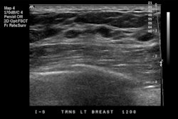
CEUS in this area can be used for preoperative sentinel lymph node identification, such as for breast cancer diagnosis. Huang added that it can optimize needle visualization for kidney diagnosis via nephrostomy insertion.
“You either have the contrast flowing into a low-pressure collecting system or the contrast gets flushed out with a high-pressure system,” he said.
Another application in this area is optimizing assessment of physiological or pathological spaces, such as for assessing volume needs for the ablation of renal cysts.
Therapeutic and future applications
CEUS can also aid in localized treatment of clots, Huang said. This involves ultrasound energy along with microbubbles within contrast agents to create a thrombolytic effect and increase cell membrane and blood-brain barrier permeability for localized drug delivery.
“The key for all these emerging applications is the precise interaction between the microbubbles and the ultrasound waves we apply,” Huang said. “With the ultrasound energy, microbubbles can rapidly change the state from what we use in a diagnostic setting as a relatively stable structure to an unstable structure.”
Additionally, Huang highlighted that CEUS can be a “perfect” modality for pregnant women and children by avoiding the use of radiation.
“It’s certainly something that’s worth thinking about,” he said. “We need more data and more applications for that, so this is something that could potentially come into place and benefit the patients we treat.”


