Identification of a toxin as the primary cause of a patient's illness is essential to prevent death or additional morbidity, and in stable cases, an MRI exam should be carried out after CT, according to researchers.
Imaging can play a central role in determining whether a toxin is present in patients and how it affects them if poisoning is known to have occurred, noted Dr. Gurubharath Ilangovan, a consultant radiologist and professor at the Tamilnadu Dr MGR Medical University, Chennai, India, and his colleagues.
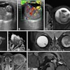
"Diverse perspectives on the indications and symptoms of common poisoning can enhance our comprehension of the fundamental mechanism, so aiding in the treatment of individuals who are acutely contaminated," they explained. "CT shows hypoattenuation bilaterally in various regions like basal ganglia, globus pallidus, and putamen based on type of poisoning. MRI gives a better resolution for cortical lesions than CT and gives better anatomical detail in the case of hemorrhage."
 Carbon monoxide poisoning. Brain MRI scan (axial FLAIR sequence) shows periventricular hyperintensities. All images courtesy of Dr Gurubharath Ilangovan et al and the ESR's EPOS database.
Imaging can help to detect early neurological changes and aid the patient in restricting the progression of the effects caused by various types of poisoning, they continued. Although toxic and metabolic brain illnesses are relatively uncommon conditions affecting the central nervous system, it is crucial to identify them because, if not treated promptly and appropriately, they can have disastrous consequences. Imaging can be used to establish the most likely diagnosis and offer prognostic data.
Organophosphate poisoning
Organophosphorus poisoning mainly results in acute neurological dysfunction and respiratory distress, the authors noted in work presented at ECR 2024. Anticholinesterases cause three distinct neurological syndromes following accidental or suicide exposure, such as life-threatening acute cholinergic crisis, intermediate syndrome with cranial nerve palsies, proximal muscular weakness, and delayed organophosphate-induced polyneuropathy.
 Organophosphate poisoning. Axial MRI scan shows fluid-attenuated inversion recovery (FLAIR) hyperintensity in caudate and lentiform nucleus of brain.
In these cases, CT shows hypoattenuation bilaterally in the basal ganglia. MRI shows diffusion restriction with symmetrical T2 or T2 fluid-attenuated inversion recovery hyperintensities involving bilateral globus pallidus with extension to the posterior limb of bilateral internal capsules and bilateral cerebellar hemispheres, which indicates acute infarcts.
Carbon monoxide (CO) poisoning
CO poisoning can have an impact on the central nervous system and the cardiovascular system. Neurological symptoms resulting from acute CO poisoning range from a minor headache to a coma and possibly death. For most patients, these symptoms can be alleviated with oxygen inhalation or hyperbaric oxygenation. Even a year after CO poisoning, very few people experience long-term neurological symptoms as a result of delayed encephalopathy.
 Carbon monoxide poisoning. Brain MRI scan (coronal FLAIR sequence) shows symmetric hyperintense foci in the globus pallidus.
Acute CO poisoning should be diagnosed and detected as soon as possible. In the absence of a history of CO exposure, early detection and diagnosis of CO poisoning can be challenging due to a lack of pathognomonic signs or symptoms.
"The delayed symptoms of CO poisoning result from the inhibition of the mitochondrial electron transport enzyme system by CO and the activation of polymorphonuclear leukocytes, which produce brain lipid peroxidation and diapedesis," the authors wrote.
In these patients, CT shows low attenuation in the globus pallidus. On MRI, the medial portions of the globus pallidus appear as bilateral areas of low signal intensity on T1-weighted images and of high signal intensity on T2-weighted and FLAIR images. The degree of carboxyhemoglobin and the range or intensity of MRI results do not appear to be related.
Methanol poisoning
Methanol is an extremely hazardous liquid that resembles ethanol in taste and smell. It is clear and colorless. It is an ingredient in a wide range of solvents that are sold commercially. Its intoxication can result from inadvertent or suicidal oral use of cleaning or antifreeze solutions, industrial solvents, or, on rare occasions, from wine or other alcoholic beverage adulteration, they noted.
Clinical manifestation varies and has a 12- to 24-hour latent period. Formaldehyde and formic acid are two hazardous byproducts of methyl alcohol metabolism that can cause a variety of clinical symptoms. Acute intoxication causes a severe metabolic acidosis that leads to severe neurological symptoms and sequelae.
"Most patients experience visual problems, such as blindness from optic nerve necrosis and demyelination, which lasts long after they recover. In the acute phase, neurological symptoms such as headaches, nausea, vomiting, dizziness, and altered sensorium are frequently experienced," they wrote.
 Methanol poisoning. Bilateral putaminal hyperintensities are visible on T2-weighted axial MRI scan of the brain.
In these patients, CT shows hypoattenuation bilaterally in the putamen. MRI shows extensive subcortical white matter and basal ganglia abnormalities consistent with edema and bilateral putamen hemorrhagic changes.
For further information about poisoning with lead, cyanide, mercury, and arsenic, please refer to the authors' full e-poster available on the ESR's EPOS website. The coauthors of the e-poster were Drs. M. Aswanth and L. Abinaya Lakshmi.