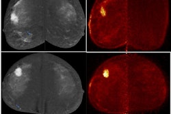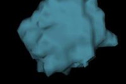
Positron emission mammography (PEM) can detect almost all breast cancer index lesions located within its scanning range without limitations due to tumor size, grade, or histological subtype, according to a Dutch study presented at RSNA 2015 in Chicago.
The findings come from an evaluation of the Mammi-PET system manufactured by OncoVision of Valencia, Spain. The technology company was established in 2003 by the University of Valencia and the Corpuscular Physics Institute. The goal was to develop a device to create high-resolution, 3D FDG-PET images of the breast in a hanging position without the need for compression.
With a viewing range of 170 mm (effective field diameter) by 180 mm (maximal length with five ring positions), the study confirmed the PEM scanner was able to visualize primary breast cancer lesions with a patient in the prone position without such compression.
"Due to its high resolution (1 mm voxel), the Mammi-PET can visualize FDG-avid lesions as small as 2mm," lead author Dr. Suzana Teixeira, PhD candidate at the Netherlands Cancer Institute at Antoni van Leeuwenhoek Hospital, told AuntMinnieEurope.com.
Patient cohort
In this study, presented by Teixeira at RSNA 2015, researchers prospectively evaluated 230 female patients with a mean age of 52 years (ranging from 24 to 82 years) and more than one histologically confirmed primary breast cancer lesion between March 2011 and March 2014. The imaging protocol included an injection of 180-240 MBq followed by whole-body PET/CT.
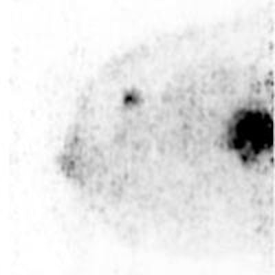
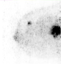 Sagittal Mammi-PET image shows two FDG-avid lesions. The smallest distal lesion, with a diameter of 2 mm, had been classified as benign (BI-RADS 2) on MRI. All images courtesy of Dr. Suzana Teixeira.
Patents received 180 MBq to 240 MBq of FDG before undergoing the PEM scan and received a follow-up standard whole-body PET/CT exam.
Researchers also evaluated all lesions discovered by the Mammi-PET with a score of 0, 1, or 2 for quantity of FDG uptake, which was tested in relation to histological results for ductal and/or lobular cancer and molecular (ER/PR/Her2) breast cancer subtype, tumor grade, breast length, maximal tumor diameter, and affected breast regions.
The PEM results then were compared with visibility scores of the primary tumor acquired using standard PET/CT.
Teixeira and colleagues scanned a total of 234 affected breasts with confirmed primary breast cancer lesions, ranging in diameter from 5 mm to 170 mm.
The Mammi-PET achieved sensitivity of 98.6% for lesions located within the device's scanning range. There were, however, 23 lesions (10%) near the pectoral muscle outside of the scanning range, which were not discovered by the device.
In the comparison with PET/CT, the PEM device detected 41 additional lesions, of which 16 abnormalities (39%) were confirmed as malignant. Fourteen lesions (34%) were visible only on the Mammi-PET.
In addition, PET/CT could not detect 15 index lesions visualized by the PEM device. Among 11 index lesions smaller than 1 cm, nine lesions (81%) were seen on the Mammi-PET.
"Of these histologically confirmed lesions, Mammi-PET was able to visualize the smallest breast cancer lesions in this series of 6 mm in diameter," Teixeira said. "Mammi-PET is able to identify heterogeneity within primary breast cancer lesions, more so than is known for PET/CT."
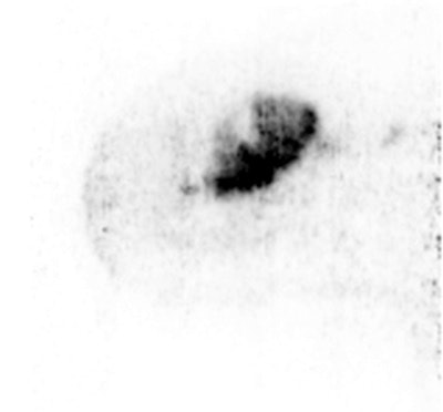 Sagittal Mammi-PET image shows one FDG-avid lesion. Inside the lesion, regions can be identified with different FDG uptake. This heterogeneity may be correlated in the future with different characteristics of the same tumor, when biopsies of the various regions are compared.
Teixeira and her and colleagues plan to continue their research by trying to identify, at least visually, heterogeneous uptake within primary cancer lesions with the development of a Mammi-PET device with an integrated FDG-guided biopsy system.
"With PET-guided biopsies, tissue can be biopsied more specifically, and opens the possibility to compare the histology of the most FDG-avid parts with those of less FDG uptake within the primary tumor," she added.
The device currently is being developed as part of a project funded by the European Commission and will be investigated for its potential clinical value in the near future.


