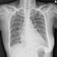
A single-exposure x-ray system allows optimization of patient radiation dose in spinal examinations, according to Spanish researchers. The device also has great potential for reducing reconstruction and motion artifacts, given the subsequent decrease of repeated images, and it may be of particular use in pediatric cases, they say.
"Children can be noncooperative patients, so one-projection studies strongly reduce movement artifacts and repetitions," noted Paula Garcia Castañon and colleagues from the Medical Physics and Radiation Protection Department at University Hospital La Princesa in Madrid. "Stitching artifacts can also be present, which definitely disappear with just one exposure."
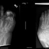
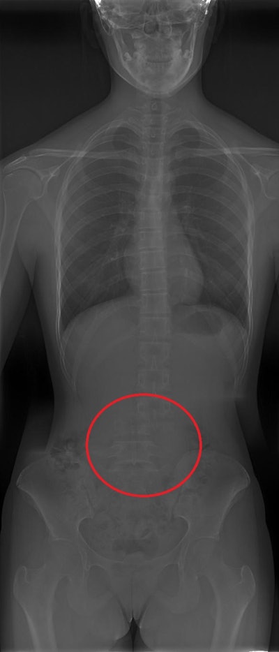 Reconstruction artifact in which a shift in part of the spine is shown. All figures courtesy of P. Garcia Castañon, S. Honorato Hernandez, C. Prieto Martin et al and ESR EPOS database.
Radiography remains the mainstay of diagnosis and evaluation of scoliosis due to its ability to image the entire spine in the standing patient to appreciate the scoliotic deformity, they explained in an e-poster presented at EuroSafe 2024. Standing radiographs allow more reliable radiographic measurements that are important in following the magnitude of the spinal deformity over time and ultimately in surgical decision-making.
The radiographic analysis of curvatures may include sideward-bending views and flexion-extension views. One anteroposterior and one lateral scoliosis x-ray images are generally performed, with systematic radiographic imaging carried out throughout the patient's course of treatment.
"During the evolution of scoliosis, a patient can be exposed to several radiographic images, so it is important to search for new techniques and or systems that produce less artifacts and which allow for an optimization in radiation exposure. This is especially important in pediatric patients, with higher radiosensitivity than adults and greater life expectancy," Garcia Castañon and colleagues added.
Two- or three-phase x-ray machines are generally used, with a digital flat panel detector that moves down the spine, obtaining different images. Usually, two to three images are performed and a reconstructed full-spine image is created, they pointed out.
An x-ray device is now available that has a detector large enough to perform full-spine images with just one exposure to the patient. The researchers sought to evaluate the performance of the one-exposure system, compared with that of a traditional machine.
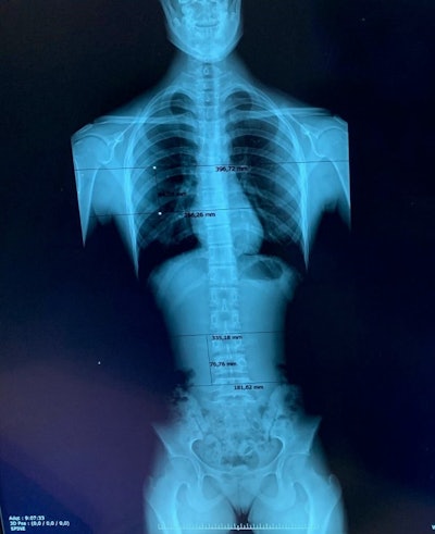 Estimated overlapping regions in the General Electric Definium 8000 unit for a patient with three projections.
Two systems are used to perform full spine studies at the Madrid hospital: a General Electric Definium 8000 machine and a Fuji FDR Smart Suspension system, which allows full-spine scans to be performed in one shot. For the latter, the detector consists of three regular detectors placed together, so there is no need to move the x-ray tube and detector down the spine, thus reducing artifacts and presumably patient effective doses due to non-overlapping.
In the study, 73 patients were imaged on the GE unit and 55 on the Fuji system. The entrance surface air kerma was calculated for each projection, from exposure and patient parameters manually recorded by the technologists during the studies. According to the images analyzed by the radiologists, a 6 cm region of overlap was considered between images for the conventional system.
Patients were categorized by age: 10-14 years (early adolescence) and 14-18 years (late adolescence). Scoliosis studies are not usually necessary in younger children.
Patients scanned on the GE unit were categorized by age and by number of images. In the 10-14 age category, 34 patients were included. 64.7% of those patients received a two-image acquisition (22 patients), and 35.3% (12 patients) were subjected to a three-image acquisition. In the 14-18 category, 70% of the patients received three images (27 patients) and 30% just two images, as the mean size of patients in this category was larger.
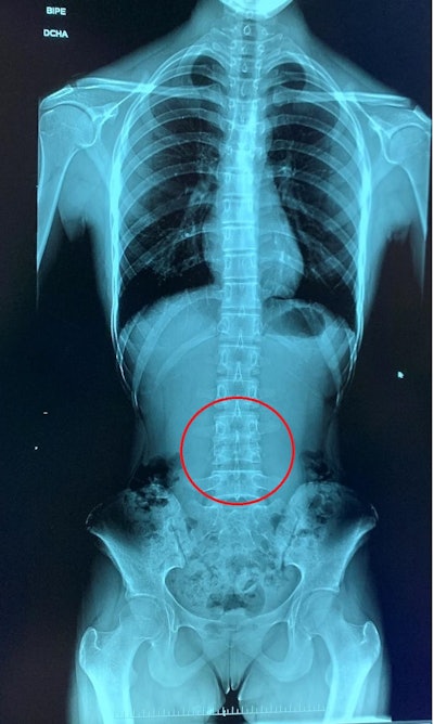 A more subtle stitching artefact is shown, where two vertebrae seem to be united.
For the first group, effective dose for two-projection studies was 60% lower than three-projection studies, while differences for the second group were less dramatic at around 30%, between larger and smaller patients from the group.
For the older patients, effective doses appeared to be lower with the one-projection unit: 3% for the two-projection group, and 34% lower when compared with the three-projection group, which constitutes 70% of the patients of this age category, the authors noted.
For younger patients, doses with one-projection unit were 14% higher when compared with the two-projection group, which was the largest sample, with 65% of the patient, and 51% lower for the three-projection group.
"Differences arise, first, from those overlapping areas needed to obtain a diagnostic image in most of the available units. On the other hand, the new unit has a direct flat panel, with no automatic exposure control, so manual technique usually will provide higher doses to an individual patient," they stated. "Further improvements of one-projection units could include automatic exposure control."
The Madrid team anticipates that an analysis of image repetition rates in both units would reinforce these promising results.
You can read the full e-poster on the EPOS database of the ESR. The work was presented at EuroSafe 2024. The coauthors were S. Honorato Hernandez, G. Paradela Díaz, C. G. Molina, P. Botella Faus, R. Rosado del Castillo, P. Chamorro, E. Garcia Esparza, C. Prieto Martin.


