Using 7-tesla MRI to image the brains of glioma patients, German researchers have correlated the protein content of those tumors with patient response to treatment and likelihood of survival, according to a study published online on 26 February in European Radiology.
The findings could be quite beneficial because approximately half of all glioma patients have an extremely aggressive form of the disease. The earlier clinicians can determine a person's responsiveness to treatment, the better they can determine the optimal course of action.
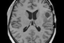
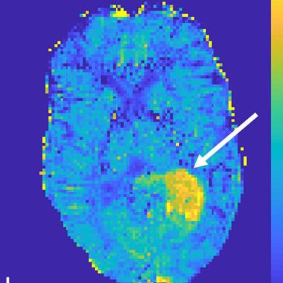
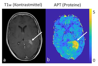 A conventional morphological contrast-enhanced 3-tesla MR image of a brain tumor (left) versus protein measurement using 7-tesla MRI (right). Images courtesy of Dr. Daniel Paech and European Radiology.
"Malignant gliomas respond very diversely to treatment," explained lead author Dr. Daniel Paech, from the division of radiology at the German Cancer Research Center (DKFZ), in a statement. "In some of the cases, postoperative radiotherapy and chemotherapy are more effective than in others. And whether the tumor has, in fact, responded to treatment cannot be told before the first follow-up care exam six weeks after treatment ends."
For this study, Paech and colleagues utilized noncontrast-enhanced 7-tesla MRI to target the chemical exchange saturation transfer (CEST) effect between the proteins and free water in tissue. Because cancer cells grow in an uncontrolled manner, they produce proteins in an equally uncontrolled manner, Paech noted.
If an ultrahigh-field MRI scan is performed, the protein signal could be used to determine how rapidly the tumor might grow. Clinicians could then choose a more effective form of therapy from the start of treatment and improve a patient's chances of recovery and/or survival.
"Our study shows that the protein signal measured in the [7-tesla] MR image is a biomarker that is associated with survival as well as with treatment response of patients: The stronger the protein signal, the poorer the prognosis," he said.
Paech and colleagues plan to continue their research with a prospective study to examine in a larger patient group whether protein measurement is possible using a 1.5- or 3-tesla MRI scanner.
"If a 3-tesla MRI machine can equally measure the elevated protein expression in the tumor, then our results may be used broadly to enhance diagnostics in glioma patients, because 3-tesla machines are available in many hospitals," he said.


