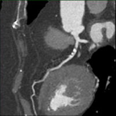
Dutch researchers are reducing the contrast dose and achieving consistent results in a coronary CT angiography (CCTA) method that tailors the scanning protocol automatically according to patient weight. The technique delivers consistent coronary artery enhancement in multiple weight categories and reduces contrast use in most patients, they report.
 Dr. Casper Mihl from Maastricht University Medical Center in the Netherlands.
In their study, Drs. Casper Mihl, Madeleine Kok, and colleagues from Maastricht University Medical Center scanned more than 250 patients in a broad range of weight categories with dual-source CCTA, individualizing contrast injection for half of the patients using contrast injection software and offering the other half of the cohort a standard contrast bolus without regard to weight. Vascular enhancement was more consistent across weight categories using automated weight-based contrast injection, andcontrast media use was reduced for most patients compared with the group receiving a standard bolus.
"Automated body-weight-adapted contrast media injection CCTA yielded comparable attenuation between different weight groups, with no significant differences in image quality, and overall contrast media volumes could be reduced in the majority of patients," Mihl said in a presentation at RSNA 2015 in Chicago.
Technology opens door to reduced contrast
Rapid technical advances in CCTA require adaptation and optimal synchronization of contrast media administration, and contrast media bolus shaping requires radiologists to adapt and synchronize the contrast administration with the scan protocol, he explained.
Enhancement of the coronary arteries is based on patient-related factors such as weight, body mass index, and heart rate, as well as scanner-based factors such as technology, volume acquisition, and pitch, and finally, factors related to the contrast itself.
Among the contrast-related factors, both the iodine delivered to the patient per second (IDR) and iodine concentration, and the amount of milligrams of iodine per milliliter (TIL) are decisive factors affecting opacification of coronary arteries, he said. Research is focused on reducing acquisition times, tube current, and contrast medium volume.
"Ideally, contrast media injection protocols should be customized to the individual patient," he said. "The aim of this study was to determine if software-tailored contrast media injections result in diagnostic vascular enhancement of the coronary arteries and if attenuation values were comparable between different weight categories."
The study included 329 consecutive patients with stable chest pain and suspected coronary artery disease who were referred for coronary CTA during a six-month period. Dual-source CT scans (Somatom Flash, Siemens Healthcare) were acquired for all studies using slice collimation of 128 x 0.6 mm, gantry rotation time of 280 msec, tube voltage 100 kV, and tube current of 320 mAs to 370 mAs.
Patients were divided into two groups. Group 1 (n = 141) received an individual triphasic bolus based on the weight category (39-59 kg, 60-74 kg, 75-94 kg, 95-109 kg) of the patient and scan duration, either high-pitch (1 sec) or dual-step prospective triggering (7 sec), with calculations performed by the contrast injection software (P3T, Bayer Healthcare).
The software used in the study allows modification of the injection rate, duration, and contrast volume for each individual patient, adapting both TIL and IDR based on a nonlinear relationship between patient weight and duration of the CT scan acquisition, according to Mihl.
Group 2 (n = 124), received a standard contrast bolus injection of 75 mL injected at a rate of 7.2 mL/sec, followed by 50 mL of saline. The tailored contrast injections given to group 1 were compared with group 2 receiving the standard injection protocol.
All patients were weighed with data entered into the system just before scanning. These triphasic bolus injections consisted of a contrast medium phase, a mixed phase with 20% contrast medium and 80% saline, and a saline phase. A test bolus was used in all patients to find the optimal start delay. For group 1, it was 20 mL of contrast at a flow rate according to P3T protocol; for group 2 it was 20 mL of contrast medium at 7.2 mL/sec. The investigators measured contrast enhancement at manually placed regions of interest in all proximal and distal coronary segments. Statistical analysis was performed using SPSS software (version 20.0, IBM).
The researchers used a test bolus at the level of the ascending aorta to assess optimal start delay (group 1: 20 mL of contrast media at a flow rate according to P3T protocol; group 2: 20 mL of contrast media at a flow rate 7.2 mL/sec). Test bolus in both groups was followed by 40 mL of saline flush at the same flow rate).
Along with quantitative measurements, an experienced cardiologist and radiologist graded the subjective scan quality of all datasets in consensus, based on a combination of the level of intravascular attenuation, the amount of image noise, and usage of a four-point grading scale, with 4 being excellent and 1 being poor.
Individualized contrast shows even attenuation across weight categories
The results showed no significant differences in all contrast enhancement values or subjective scan quality between the two groups (p = 0.311). However, attenuation was far more homogeneous for the software-tailored patients in group 1, Mihl said.
In group 1, enhancement was sufficient in all weight groups with values of 325 HU or greater, and there were no significant differences in mean attenuation values between different weight classes (p > 0.173). Proximal and distal values, respectively, were as follows:
| Group 1: Coronary artery attenuation in proximal and distal coronary regions | ||
| Weight range | Proximal values (HU) | Distal values (HU) |
| 39-59 kg | 449 ± 65 | 373 ± 58 |
| 60-74 kg | 443 ± 69 | 367 ± 81 |
| 75-94 kg | 427 ± 59 | 370 ± 61 |
| 95-109 kg | 427 ± 73 | 347 ± 61 |
 Chart shows even and adequate attenuation with individually tailored contrast across multiple weight categories and two scanning protocols. Image courtesy of Dr. Casper Mihl.
The attenuation was uniformly strong in group 1, and in none of the vascular segments or weight classes did mean attenuation fall under 325 HU. Moreover, "standard deviation was lowest in group 1, indicating that attenuation was more homogeneous among patients of this group," he said.
On the other hand, group 2 results showed "significantly lower attenuation levels in higher weight groups, while slender patients showed markedly increased enhancement levels," Mihl said. Also in the control group, attenuation values between proximal and distal segments in the control group decreased significantly (p < 0.008) within higher weight classes and mean attenuation of the distal left anterior descending (LAD) for patients weighing 95-109 kg did not reach diagnostic attenuation values at 305 ± 35 HU.
Finally, the use of automated injection protocol reduced the contrast media dose for most patients compared with the fixed-dose protocol.
| Contrast volume by patient weight | |
| Mean contrast volume (mL) | |
| Group 1 (P3T) | |
| 39-59 kg | 54 ± 6 (p < 0.001) |
| 60-74 kg | 61 ± 7 (p < 0.001) |
| 75-94 kg | 71 ± 8 (p < 0.001) |
| Group 2 (control) | 75 ± 0.1 |
"Individually tailored contrast media injection protocols yield diagnostic attenuation in all scans and a more homogeneous enhancement pattern between different weight groups compared with a fixed injection protocol," Mihl stated. "In addition, overall contrast media volumes could be reduced for the majority of patients utilizing the P3T software."
Individually tailored contrast media injection protocols in CCTA also allow for substantial reductions in contrast volume for most patients, while maintaining diagnostically adequate images, he said.


