Upper gastrointestinal radiography and CT are vital to assess complications after bariatric surgery, but reading these examinations is not easy and understanding the surgical techniques is essential, French researchers have found.
The main abdominal complications are anastomotic leak and small-bowel occlusions, Dr. Catherine Ridereau-Zins, from Angers University Hospital, told delegates at last month's European Society of Gastrointestinal and Abdominal Radiology (ESGAR) meeting in Barcelona.
"Gastrojejunal (GJ) anatomosis leak is one of the worst early complications requiring revision surgery in an emergency," she explained. "Fluid collection near the GJ anastomosis suggests a fistula of the anatomosis. A fistula in the remnant stomach due to disruption of the gastric staple line -- mechanical failure of staples -- may be responsible for weight gain. Small-bowel occlusions, including adhesions and internal hernia, may occur due to the prolapse of small-bowel loops into mesenteric defects."
Obesity is a major public health challenge due to serious cardiovascular, metabolic, and pulmonary risk factors, according to Ridereau-Zins. The first treatment is medical management via a balanced diet, physical fitness, and behavioral therapies, but the use of bariatric surgery has increased for patients with morbid obesity. The most common procedures are adjustable gastric banding, Roux-en-Y gastric bypass, and sleeve gastrectomy, all of which are performed laparoscopically.
In addition to anastomotic leak, early complications may be a fistula, abscess, splenic or left hepatic lobe infarction, hematoma, gastrointestinal bleeding, and general complications such as pulmonary embolism and small-bowel occlusion. Late complications include small-bowel occlusion, adhesions, internal hernia, and migration of the gastric banding.
The upper gastrointestinal (GI) contrast study can help depict complications. It should be performed two days postoperatively when there is a clinical suspicion of a fistula and at one-year follow-up to analyze morphology of the GI tract.
"Initial imaging is performed with a water-soluble contrast material to assess for anastomotic leak. Later a barium sulfate suspension is used to analyze the surgical results with a better contrast," Ridereau-Zins stated. "The exam begins with a plain film to help identify surgical material. Then radiographs are performed after the patient swallows a high quantity of contrast material (100 mL)."
Reading the images can be difficult, especially in the postoperative period, due to bad contrast for these large patients with a water-soluble contrast material, dilatation of the esophagus, size of the neo pouch, permeability of the anastomosis, and opacification of the initial small bowel. A CT exam should be performed when an upper GI contrast study has failed in a suspected case of fistula, and it can also rule out other complications.
"As usual in abdominal CT, it is helpful to follow the GI tract from the esophagus to the ileum, especially in coronal views. But the key is understanding the surgical procedure and knowing normal postoperative findings for each surgical technique," she noted.
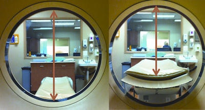 Though CT aperture diameters may be accurate in the horizontal plane, they do not account for the table thickness entering the gantry bore. The vertical diameter is actually 20 cm less than the published diameters, limiting its use in morbidly obese patients. All images courtesy of Dr. Rajeev Suri, an associate professor of vascular and interventional radiology at the University of Texas Health Science Center in San Antonio, U.S.
In an e-poster presented at ESGAR 2013, Dr. Rajeev Suri, an associate professor of vascular and interventional radiology at the University of Texas Health Science Center in San Antonio, U.S., elaborated on the logistical difficulties posed by the growing obesity epidemic. These include the weight limits and aperture diameters of imaging equipment, scheduling and transport issues, working out how well the patient will fit in the imaging equipment, and modification of equipment settings to optimize image quality for the obese patient.
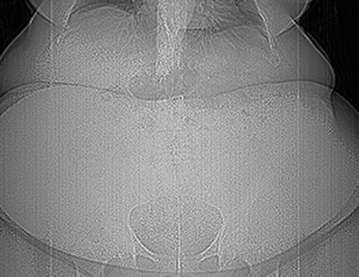 Digital radiograph of a 635-lb patient after a motor vehicle accident. To image this patient with CT, knowing the weight limits of the available CT scanner is critical -- most CT scanners have a weight limit of 450 lbs to 500 lbs. CT scanners available with higher weight limits include: Toshiba Medical Systems (660 lb, Aquilon One); Philips Healthcare (650 lb: Brilliance Big Bore); GE Healthcare (650 lb: Lightspeed Xtra), and Siemens Healthcare (660 lb: Somatom Definition).
"Current imaging technology is limited in providing the desired quality of imaging in obese patients," he wrote. "Radiologists and technologists need to be aware of the limitations of imaging equipment and the equipment adjustments that can be made to improve image quality in obese patients. Best technique depends MORE on weight limit and aperture sizes THAN the appropriateness of the exam. Consider portable radiography or sonography equipment for patients who cannot be transported."


