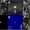
MRI tends to be the best modality for investigating patients undertaking sex reassignment surgery (SRS) because it allows a detailed assessment of the new pelvic anatomy after male-to-female and female-to-male SRS, according to award-winning research from Italy. Furthermore, it identifies postoperative complications and provides information that can be useful in planning further intervention.
Color Doppler ultrasound, however, still has a role in assessing vascularity of the neophallus and in guiding interventional procedures, and CT angiography is the state-of-the-art for preoperative vascular mapping in microsurgical free-flap phalloplasty. In female-to-male transsexuals, urethrography allows full evaluation of the neourethra, noted Dr. Michele Bertolotto and colleagues from the department of radiology at the University of Trieste.
"Sex reassignment surgery is a complex and difficult surgery that is fraught with risk," they noted in an e-poster that received a certificate of merit at RSNA 2012 in Chicago. "Optimal treatment of transsexual patients requires a multidisciplinary [team] with a nucleus of dedicated physicians. Radiologists must be confident with the pelvic anatomy after genital reconfiguration and with possible postoperative complications."
Many surgical approaches exist, especially for female-to-male SRS, and all have pros and cons as well as risks. Imaging may provide information to help the surgeon adopt the most suitable operation for each patient, they explained.
Male-to-female SRS requires orchidectomy, extirpation of the corpus cavernosum, shortening of the urethra, creation of a sensitive neoclitoris and of the neovagina, mons pubis, and vulva with labial structures. Female-to-male SRS requires hysteroannessiectomy, mastectomy, and phalloplasty using various techniques. A second operation is usually done when sensitivity is present in the neophallus to insert testicle implants and a semirigid/inflatable penile prosthesis.
In the pelvis of a male-to-female transsexual, full evaluation of the normal postoperative changes and presence of postoperative complications can be made with MRI, the authors stated. T2-weighted images are obtained, at least in the sagittal and axial plane, followed in some cases by T1-weighted fat-suppressed images in the same planes before and after gadolinium contrast administration. T1-weighted and proton density-weighted images are less commonly used. To better evaluate the neovagina, they recommend distension gel and introduction of an inflatable device.
"The cavity for the neovagina is obtained with wide blunt dissection of the space anterior to the rectum and posterior to the prostate. This is the most dangerous phase of the operation because accidental rectal injuries are possible. MRI provides an accurate evaluation of the thickness of the recto-neovaginal septum which is only a few millimeters," they noted.
The bulbo-cavernous muscle may be used to reinforce the distal portion of the septum, and is best identified to the lower portion of the neovagina on T1- and proton density-weighted axial and sagittal high-resolution images.
MRI allows excellent evaluation of the neoclitoris, according to the researchers. The spatulated urethral stump, glans remnant, and neurovascular bundle are best evaluated on T2-weighted and gadolinium-enhanced T1-weighted images; the latter allows evaluation of vascularization. Complications such as blood extravasation and ischemic changes can be identified.
Compared with ultrasound, MRI provides a more panoramic view for planning of interventional and surgical procedures, according to the authors. The best depiction of cavernosal and spongiosal remnants and of their relationship with the surrounding structures is obtained on T2-weighted images after a prostaglandin E1 injection.
A minor but relatively common postoperative complication is neovaginal prolapse. Bleeding most commonly occurs around the neoclitoris, which is highly vascularized, they concluded.


