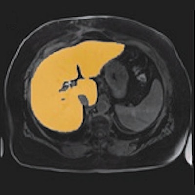
Modern machine-learning algorithms should not replace radiologists but should support them in their everyday work. A successful example of this is the deep-learning algorithm developed by two German radiologists to assist with liver MRI scans. The procedure can also be applied to other diagnostic issues.
Today, radiologists are increasingly required to deliver 2D cross-sectional images and also provide 3D datasets. For example, surgeons performing operations in the abdomen often want to know the exact details of the spatial structure of an organ and the adjacent regions to enable optimal surgery. Cardiologists and heart surgeons who implant artificial heart valves also expect 3D analysis.
 The graphic shows the segmentations of an MRI scan (A) with the manual (B) and the automatic variant (C). Panel D illustrates the comparison between manual and fully automatic segmentation. The green pixels indicate the areas of concordance, while the red pixels represent the areas of deviation. Image courtesy of Dr. Niklas Verloh.
Many of these 3D analyses require segmentation of the organs. Organs are subdivided into individual anatomical or functional sections to provide the requesting physician with the desired information. This segmentation takes a lot of time if the radiologist has to manually do it. However, CT software programs are now available, which can at least partially automate segmentation.
Liver segmentation: A real challenge
 Dr. Niklas Verloh. Image courtesy of the DRG.
The task is somewhat more complicated in MRI. "This, in particular, applies to the liver, where manual segmentation of the MRI dataset takes about 10 to 20 minutes, which is very time-consuming. The shape of the liver can be influenced by both respiration and various diseases. This makes it difficult to analyze," emphasizes Dr. Niklas Verloh from the Institute for X-Ray Diagnostics at University Hospital Regensburg.
Difficult but not impossible: Two years ago at a workshop from the series Researchers for the Future, organized by the German Radiological Society (DRG), Verloh met his colleague Dr. Hinrich Winther of the Institute for Diagnostic and Interventional Radiology at Hannover Medical School. Together, they decided that the times of manual segmentation of 3D MRI scans of the liver slowly needed to come to an end.
"Modern machine-learning approaches have made these more complex analyses possible. There are now neural networks based on 3D datasets that provide significantly better results than older neural networks," Verloh said. The two radiologists from Regensburg and Hannover used such a neural network, which they trained and validated with a total of 100 patients in a multistage procedure.
The segmentation is completed within a minute. The validation consequently shows that the trained algorithm can manage the segmentation of the liver with high accuracy using the MRI dataset.
"In terms of the differences between the evaluations, we are currently at a stage where they are comparable to the differences between two human evaluators. This is a very good result at such an early stage of training," Verloh noted.
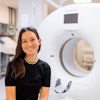
 Dr. Hinrich Winther. Image courtesy of the DRG.
In fact, the radiologists looked at some segmentations in detail and found something interesting. The differences to human findings are often due to the fact that the algorithm has segmented more precisely than humans. The question, therefore, arises as to whether the evaluation by a radiologist can really be regarded as the gold standard.
"In theory, a person can segment 100% accurately, but, due to the time-consuming nature of the activity, this is only possible to a limited extent in routine clinical work," Verloh added. The algorithm, on the other hand, is very fast: In an MRI scan with 64 layers, the neural network requires an average of 60 seconds for segmentation.
Further application scenarios are already being tested.
What works for the liver can, of course, also work for other organs. At the Hannover Medical School, Winther has already used the segmentation algorithm to segment the lungs and the heart. "The first experiences here are also very promising," he explained.
However, it is clear that all application scenarios of the segmentation algorithm are currently still research projects: "These are not yet approved medical products and are, therefore, not tools for everyday clinical use at the moment. This would be the next step, for which technical partners are required," Winther concluded.
Further details will be available at the following session at the 100th German Radiology Congress:
WISS 205 -- Artificial Intelligence on Thursday, 30 May 2019, from 14:00 to 15:40, Hellmann room
- 14:00 to 14:10 -- Use of a 3D Neural Network for Liver Volume Determination in 3T MRI by Dr. Niklas Verloh
- 14:40 to 14:50 -- Deep Semantic Segmentation of 4D DCE MRI Lung Scans to Assess Clinical Biomarkers in Chronic Obstructive Pulmonary Disease by Dr. Hinrich Winther
Editor's note: This is an edited version of a translation of an article published in German online by the German Radiological Society (DRG, Deutsche Röntgengesellschaft).
