The European Council took a significant step forward in advancing women's health when it endorsed digital breast tomosynthesis (DBT) as the new standard for breast cancer screening in December 2022. This revision of the screening guidelines was made because of the growing evidence that supports it as a superior screening modality, such as the Tomosynthesis plus Synthesized Mammography (TOSYMA) study that found DBT plus synthesized mammography detected 3.5 times more invasive cancers than digital mammography alone in the first screening round.1
 Dr. Jacopo Nori.
This technology is a powerful tool in the detection of breast cancer during primary screenings. With the recent evolution of breast cancer screenings, contrast-enhanced mammography (CEM) has emerged as a promising supplemental imaging technology that works alongside DBT to further improve patient outcomes. It has the potential to significantly improve detection rates, but education and awareness remain a significant barrier to its adoption.
Challenges to widespread CEM adoption
CEM is a relatively new technology compared with established methods like MRI, and at Careggi University Hospital, this has led to a knowledge gap among some radiologists. We began using CEM in 2016 for approximately 10 exams per week; now we do 10 exams per day with the modality. While our use of the technology has increased over the years, we have found that radiologist knowledge about CEM has not kept pace. Radiologists joining our department simply are not educated or experienced with it, even though research shows that it is a quicker alternative to MRI.2
CEM pairs 2D and tomosynthesis images, all under one compression, providing anatomical and functional imaging in one exam.3 The modality leverages a contrast agent, similar to what is used in CT technology, and it accumulates where lesions are forming and growing. For radiologists unfamiliar with CEM, the resulting images can present as a challenge, requiring them to effectively integrate a variety of data points across multiple images for accurate diagnosis. They must acclimate to this new workflow.
For those who have started using CEM at Careggi University Hospital, the feedback has been very positive. The quick learning curve has facilitated a seamless integration of this modality, and we find that young doctors can approach the technology quite easily. After an initial learning period, users report that the system is easy to navigate.
Although it may take a little time to get fully accustomed to using the technology and integrating it into the breast screening pathway, there are significant benefits to CEM. One is that it can help significantly improve detection rates when you compare it to digital mammography alone, as the modality reveals hidden information that standard tests often miss.4
At Careggi University Hospital, we have found it to be highly beneficial as a diagnostic tool because the extra information helps us plan surgeries better. In fact, CEM has helped us discover unsuspected issues in approximately 23% of cases at our breast imaging facility, potentially altering the surgery plan for the betterment of the patient.
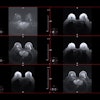
 Clinical patient images comparing details available on a 2D mammography system (left) with contrast-enhanced software (center) and on a 3D mammography gantry (right). Figure courtesy of Hologic.
Education remains key to CEM adoption. Once the initial training investment is made, it can help simplify supplemental screenings and reduce dependency on MRI. This could help streamline the diagnostic process for patients.
Benefits of CEM vs MRI: Collaboration, not competition
Rather than viewing MRI and CEM as competitors, it is important to recognize their respective strengths. CEM complements MRI – it doesn't replace it. Studies show that CEM and breast MRI both have comparably high sensitivity in the detection of breast lesions, 5 meaning CEM can offer a faster alternative to MRI without compromising on results.6 This enables facilities to reserve MRI for when it is needed and reduce delays between screening and diagnosis for breast cancer patients. While MRI scans produce beautiful images, their analysis typically requires more time,6 making CEM a more suitable choice for many breast patients.
Despite its many advantages, the biggest barrier to wider adoption of CEM is a lack of awareness and training among radiologists. To address this, we need more widespread education and implementation to make radiologists acquainted with the benefits and help them become more acclimated to CEM.
This technology enhances detection, is quick to learn, enables rapid diagnoses, promotes communication with surgeons, and helps centers manage their resources efficiently. By implementing educational programs, increasing awareness through conferences and publications, and integrating CEM into the latest mammography units, we can unlock the full potential of this technology to improve early breast cancer detection and patient outcomes.
Dr. Jacopo Nori is chief of breast imaging at Careggi University Hospital, Florence, Italy.
The comments and observations expressed herein do not necessarily reflect the opinions of AuntMinnieEurope.com, nor should they be construed as an endorsement or admonishment of any particular vendor, analyst, industry consultant, or consulting group.
REFERENCES
- Weigel S, Heindel W, Hence H-W, et al.: Breast Density and Breast Cancer Screening with Digital Breast Tomosynthesis: A TOSYMA Trial Subanalysis. Radiology, 2023;306(2):e221006.
- Li L, Roth R, Germaine P, et al, Contrast-enhanced spectral mammography (CESM) versus breast magnetic resonance imaging (MRI): A retrospective comparison in 66 breast lesions. Diagn Interv Imaging 2017 (February);98(2):113-123.
- Burhenne LJW, Wood SA, D'Orsi CJ, et al. Potential Contribution of Computer-aided Detection to the Sensitivity of Screening Mammography, Radiology 2000; 215:554-562
- Coffey K, Jochelson MS. Contrast-enhanced mammography in breast cancer screening. Eur J Radiol. 2022 Nov;156:110513.
- Gelardi F, Ragaini EM, Sollini M, et al. Contrast-Enhanced Mammography versus Breast Magnetic Resonance Imaging: A Systematic Review and Meta-Analysis. Diagnostics (Basel), 2022 Aug 4;12(8):1890.
- Hobbes M, Taylor D, Buzynski S, et al. Contrast-enhanced spectral mammography (CESM) and contrast enhanced MRI (CEMRI): Patient preference and tolerance. J Med Imaging Radiat Oncol. 2015 Jun;59(3):300-5.