Radiologists and trainees must become more familiar with different radiological signs to accurately interpret breast images and provide an accurate diagnosis. That’s the view of award-winning researchers.
“Identification of different radiological signs is critical for early diagnosis and effective treatment of various medical conditions, including breast cancer,” noted Dr. Karina Pesce, PhD, and colleagues from Hospital Italiano de Buenos Aires in Argentina. “Knowledge of clinical and radiological signs in breast imaging can help prevent diagnostic errors and reduce the number of unnecessary medical tests.”
The semiotic description of images, including signs, their reading and interpretation, holds significant value for radiology residents in training, and it facilitates a practical approach to images and provides a structured diagnostic framework for identifying findings, they explained in an RSNA 2023 poster that received a certificate of merit.
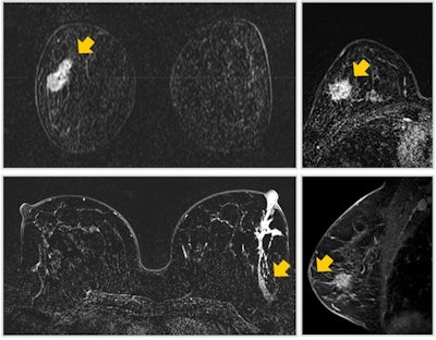
 Comet sign: A lesion with enhancement characteristics that typically exhibits a "tail" that extends into the parenchyma, frequently oriented toward the nipple, resembling the tail of a comet. When a "comet tail" emerges from an enhanced lesion, it serves as a robust indicator of early ductal carcinoma in situ, particularly when it points toward the nipple or aligns with the ductal system. Less probable scenarios, which can be readily discerned from the patient's medical history, include previous biopsies or surgeries in the preceding months. All images courtesy of Dr. Karina Pesce, PhD, and presented at RSNA 2023.
The tent sign in mammography describes the appearance of the breast parenchyma’s posterior edge when a mass causes it to retract, forming an inverted “V” that resembles a circus tent tip, according to the authors.
A halo sign in mammography comprises a complete or partial radiolucent ring surrounding the periphery of a breast mass. “While the presence of the sign guides us towards benign pathology, we must not forget that it can also be exhibited by other malignant entities such as triple-negative basaloid breast carcinoma or papillary carcinoma.” To guide the diagnosis, compare the image with previous studies, consider the patient’s clinical data, and supplement with ultrasound, they recommend.
The tattoo sign in mammography appears as calcifications that maintain a fixed and reproducible relationship to one another on mammograms obtained with similar projections at a different time. Consistent calcification patterns that maintain a fixed spatial relationship can indicate a dermal origin, and recognizing the tattoo sign can help reduce the need for unnecessary needle localization and biopsy procedures, they stated.
The black star sign consists of a central radiolucent core with long, thin, radiating spicules arranged parallel to radiolucent linear structures. Variability of appearance in different projections and the absence of palpable lesions/skin changes are other features to look for. “The black star sign indicates radial scar diagnosis, but not all radial scars look like this; some have a radiodense center called a white star, like carcinoma.”
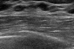
The picket fence sign is seen when many closely spaced Cooper’s ligaments are involved in cancer, creating a shadowing area resembling a picket fence profile. The Golden Gate sign occurs when two or three adjacent Cooper’s ligaments are affected by cancer, creating a hypoechoic area resembling a suspension bridge in profile.
The Mickey Mouse sign indicates metastatic invasion of subcapsular and cortical sinuses, and it is eccentric, giving the characteristic eccentrically enlarged cortex. The rat bite sign consists of a hilar indentation with a convex appearance.
In B-mode ultrasound, the yin and yang sign presents as an oval or round nodular image with a clear anechoic border. Without color Doppler, it might be confused with a cyst. Color Doppler ensures precise diagnosis; you see a mix of red and blue colors, indicating blood circulation and forming the yin and yang sign. When performing a breast ultrasound, don’t forget to ask the patient if she has recently had an interventional breast procedure, the authors noted.
In the onion ring sign, an epidermoid cyst of breast skin gives an onion ring appearance of alternating concentric hyperechoic and hypoechoic rings on sonography, representing multiple layers of keratin. The importance of recognizing this lesion lies in its potential to mimic any benign or malignant breast lesion, both clinically and radiologically.
The snowstorm sign on ultrasound comprises an echogenic mass with posterior shadowing – a “snowstorm” appearance of free silicone. Possible causes are bleeding, silicone injections, and implant rupture.
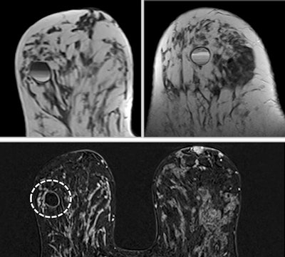
 Fat-fluid levels sign: T1- and T2-weighted MRI shows cyst with thin septa, heterogeneous content, and fat-fluid levels, which is compatible with fat content, suggestive of galactocele. A cystic mass with a fat-fluid level is a diagnostic indicator of a galactocele, with the fat content above and the water content remaining at the bottom.
On MRI, the four signs of breast implant rupture are the linguine sign (an elastometric shell of a ruptured implant floating within silicone gel), the keyhole sign (focal silicone invagination between the inner shell and fibrous capsula), the salad oil drop sign (silicone gel mixed with reactive subcapsular fluid), and the subcapsular lines sign (hypointense lines surrounded by silicone gel that run parallel to the fibrous capsule). If only the salad oil drop sign is visualized, it is impossible to conclude that the implant exhibits an intracapsular rupture, they wrote.
The donut sign comprises a small, clearly defined ring that maintains its sharp outline throughout the dynamic study and does not demonstrate “filling in.” It is not indicative of carcinoma and is more likely to be a well-defined benign lesion, typically a papilloma or fibroadenoma.
Carcinoma may also exhibit ring enhancement, but this is typically observed only in the initial dynamic sequence; subsequently, the enhancement gradually fills in the central portion of the lesion, resulting in a relatively uniform enhancement of the entire mass, according to the researchers.
A continuous vessel sign on breast MRI occurs when a hyperintense spot occupies a similar position in neighboring slices and can be tracked from one slice to the next along its course. It is important to recognize this sign so as not to mistake a vessel with a small mass or carcinoma, they added.
A fascial tail sign involves the identification of a predominantly low-to-intermediate signal intensity mass with linear extensions along the superficial fascia at the margins and low-signal intensity bands. The fascial tail appears as a tapering marginal extension of the tumor along the fascial plane. The tail enhances after contrast media administration, confirming fibromatosis of the breast.
Breast within a breast sign on mammography is considered pathognomonic of breast hamartomas. The imaging appearance of these uncommon benign tumors can vary widely.
An eclipse sign indicates sharply demarcated enhancement around a cyst, resembling a solar eclipse. The halo observed in inflammatory cysts is noticeable in late dynamic MRI sequences, while the typical annular enhancement of breast carcinoma appears in early sequences and persists into the late ones. The halo is a thin and homogeneous ring, whereas the annular enhancement in carcinomas is thick and heterogeneous. In T2-weighted sequences, if it’s a cyst, it tends to be uniformly hyperintense, in contrast to a carcinoma, which exhibits heterogeneous hyperintensity, the authors wrote.
A black hole sign has been described as another characteristic sign on MRI to help diagnose fat necrosis, consisting of marked central hypointensity of the lesion on short-tau inversion recovery (STIR) images when compared with surrounding fat.
Others to bear in mind are the necrosis sign (invasive carcinomas promote tumor growth by inducing new blood vessel formation and neoangiogenesis), the blooming sign (a lesion with initially sharply shaped borders one minute after bolus injection and becoming blurred seven minutes after bolus injection, with the lesion showing rapid enhancement during the first two minutes after bolus injection), the adjacent vessel sign (axial and coronal maximum intensity projection views allow a comparative evaluation of symmetry or asymmetry, and 3D rotation on a workstation allows a detailed evaluation of the surrounding vessels and a lesion), and the hook sign (a fine, threadlike connection passing from the lesion to the pectoralis muscle).
The co-authors of the RSNA exhibit were Dr. Ricardo Anibal Sinner I, Dr. Giannina Maria Secco, Dr. Maria Paz Swiecicki, and Dr. Maria Jose Chico.


