A new whole-brain MRI technique called field-cycling imaging (FCI) identifies subacute ischemic stroke at field strengths as low as 0.2 millitesla (mT), according to a study published on 27 August in Radiology.
The technique could make MRI more accessible, wrote a team led by Vasiliki Mallikourti, PhD, of the University of Aberdeen in the U.K.
"The inequity in access to detailed stroke imaging highlights a potential role for dedicated ultralow-field-strength (< 0.2 tesla) scanners, which can be used to rapidly and safely to detect ischemic stroke," the group noted. "Siting these in ambulances or rural settings could help direct initial or follow-up care."
Patients presenting with stroke need rapid diagnosis, and researchers have continued to develop imaging modalities that don't impart high levels of radiation and, in the case of MRI, have low magnetic field strength. FCI measures "change in T1 relaxation time constant of tissues over a range of low magnetic field strengths" (0.2 millitesla [mT] to 0.2 tesla) by switching between different fields during the pulse sequence, thus offering "new sources of contrast, including some invisible to clinical MRI scanners," Mallikourti and colleagues wrote. The technology was developed at the university.
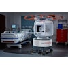
 Photograph of the field-cycling imaging (FCI) scanner prototype, with the head coil in place. Image and caption courtesy of the RSNA.
The investigators conducted a proof-of-concept study that explored whether a prototype whole-body FCI scanner could identify infarct areas in patients with subacute ischemic stroke. The research included nine participants with confirmed stroke who were admitted to a single-center stroke unit between February 2018 and March 2020 and April 2021 and December 2021; patients underwent FCI between one and six days after the event. The FCI exams were taken at four to six fields between 0.2 mT and 0.2 tesla with five evolution times ranging from five to 546 milliseconds. The researchers generated T1 maps and two readers blinded to the clinical images rated the FCI scans.
Mallikourti and colleagues reported the following:
- FCI scans below 0.2 tesla exhibited hyperintense T1 regions that corresponded to the infarct region identified at baseline imaging and that were visually confirmed with 86% interrater agreement.
- The infarct-to-contralateral-tissue contrast ratio increased as magnetic field decreased between 0.2 mT and 0.2 tesla (p < 0.001).
- T1 dispersion slopes differed between infarct and unaffected tissues (median, 0.23 versus 0.35; p = 0.03).
 T1 maps generated after processing the field-cycling imaging scans in a 79-year-old male participant with right occipital lobe infarct. Images were processed with a total generalized variation regularization. The infarct, shown in yellow, demonstrates higher T1 relaxation time constant than the unaffected brain tissue. The infarct is clearly visible at 21.1 and 2.2 mT, where the infarct to contralateral tissue contrast ratio is higher (percentage difference in T1 of 12.3% at 200 mT, 46.6% at 21.1 mT, and 46.2% at 2.2 mT). The color bars indicate the T1 values in milliseconds. Images and caption courtesy of the RSNA.
The study findings are promising, according to the authors.
"The ability to safely measure T1 relaxation time constant changes due to cellular-level alterations at very low magnetic field strengths could prove a useful and safe adjunct in assessment and follow-up of patients with stroke, particularly in rural or low-resource settings," they concluded.
The complete study can be found here.