Patient positioning is critical to detect breast cancer successfully when using PET/MRI or MRI alone, and placing patients in the prone position greatly increases the chances of discovering lesions compared with when they are in the supine position, according to German research presented at the recent RSNA 2017 meeting.
"Breast cancer is one of the leading causes of death in women worldwide. Therefore, primary tumor staging can provide an efficient and proper treatment strategy," said study co-author Dr. Johannes Grueneisen from Essen University Hospital. "Whole-body MRI and whole-body PET/MRI in the supine position is not very sufficient for primary tumor detection and evaluation."
Using PET/MRI as a one-stop imaging tool, which would be desirable for efficient local tumor staging and potentially more accurate than conventional staging, would be time-saving and would increase patient comfort, he added.
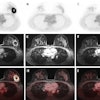
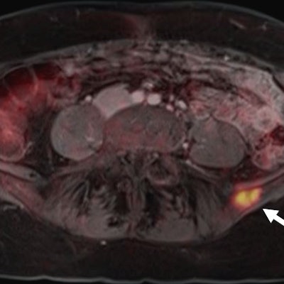
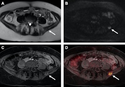 Images are from the January 2017 study of a patient with recurrent breast cancer and a PET-positive bone metastasis of the left iliac wing, which can be identified on T2-weighted half-Fourier acquisition single-shot turbo spin-echo (HASTE) (A) as well as on diffusion-weighted imaging (DWI) (B, arrows). The identical tumor lesion shows moderate contrast-enhancement on T1-weighted volumetric interpolated breath-hold examination (VIBE) images (C) and can be detected with a higher conspicuity after image fusion on PET/MRI (D). All images courtesy of Dr. Johannes Grueneisen.
Study logistics
The researchers looked at 32 patients (31 women, 1 man; mean age of 38) with histopathologically confirmed breast cancer. Each patient underwent a whole-body FDG-PET/MRI exam on an integrated scanner featuring a 3-tesla MRI system (Biograph mMR, Siemens Healthineers).
The imaging protocol included a breast PET/MRI scan with the patient in the prone position. The patient then was repositioned to the supine position. Finally, an additional whole-body MRI scan was performed.
Two radiologists with experience in hybrid imaging and breast MRI initially analyzed the hybrid and MR image sets to evaluate the primary tumors. A second reading session followed for the whole-body MR images and the combined whole-body PET/MR images for primary tumor evaluation as well as the detection of potential recurrence of lymph nodes or distant metastases.
Position comparisons
In reviewing the results, the researchers found that dedicated MRI and PET/MRI scans taken in the prone position correctly identified all 32 patients with breast cancer (100%). By comparison, in the supine position, breast MRI correctly identified 28 patients (87%) with breast cancer, compared with supine breast FDG-PET/MRI, which correctly identified 29 (90%) cancer patients.
"In the lesion-based analysis, we focused on how many primary tumor lesions could be seen," Grueneisen said. "Breast MRI and breast PET/MRI were the same in the prone position, but whole-body MRI in the supine position detected fewer primary tumor lesions than whole-body PET/MRI."
Based on the reference standard of 51 lesions, both MRI alone and PET/MRI discovered all 51 lesions (100%) with patients in the prone position, with two false-positive findings.
In the supine position, breast MRI detected 36 (70%) of the 51 lesions with five additional false-positive results. Breast FDG-PET/MRI in the supine position performed slightly better with 44 correctly discovered lesions (86%) and six additional false-positive findings.
The researchers also looked at the modalities' performances for multifocal and multicentric lesions. While breast MRI and a breast PET/MRI performed equally well in the prone position, the modalities did not recognize multifocal disease in two cases in the supine position. In addition, breast MRI did not detect multicentric disease in three cases, compared with breast PET/MRI, which missed one multicentric lesion.
"Dedicated breast imaging in prone positioning with or without FDG information is superior to examinations in supine position," Grueneisen and colleagues wrote in their abstract."While FDG information did not improve the diagnostic value of dedicated prone breast MRI, adding FDG information to supine breast examination led to an increase of tumor detection, but also of false-positive findings."
Researchers' track record
Grueneisen and his fellow researchers have investigated the use of PET/MRI for women's imaging for several years. In a January 2017 study, they found the hybrid modality achieves "superior restaging" with breast cancer patients compared with MRI alone.
The paper followed a 2015 study in which the group found PET/MRI achieved high diagnostic performance for restaging gynecological cancer patients compared with FDG-PET/CT, with only a slightly longer scan time and "markedly reduced" radiation exposure. In 2014, the German researchers concluded that adding diffusion-weighted imaging (DWI) to FDG-PET/MRI to stage women with primary or recurrent pelvic malignancies contributed minimal value and is not worth the additional scan time.

