Medical physicists from Nantes, France, have generated a series of medical isotopes from the irradiation of thorium-232 (Th-232). One of the products decays into thorium-226, a promising isotope for alpha radionuclide therapy, while another product is molybdenum-99 (Mo-99) -- an increasingly sought-after isotope used in diagnostics.
Radioisotopes are used in medicine for both cancer therapy and diagnosis. For therapy, there is a small demand for alpha emitters -- isotopes that decay with the release of a helium nucleus -- because these can deliver a high dose of radiation over a very short range. For diagnosis, there is a sharply increasing demand for metastable technetium-99 (Tc-99m), a gamma-emitting isotope that is already used in upwards of 25 million examinations every year.
Tc-99m is the decay product of Mo-99, which is generated by five aging reactors across the globe -- HFR in the Netherlands, SAFARI-1 in South Africa, BR2 in Belgium, NRU in Canada, and OSIRIS in France. NRU and OSIRIS, which together deliver nearly half of the world's Mo-99, are expected to cease production over the next two years, prompting several medical authorities to warn of a looming "Tc-99m crisis" -- although new reactors are due to come online in France and Australia.
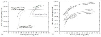
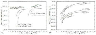
Production yields of activation (a) and fission products (b).
With the demand for alpha emitters and Mo-99 in mind, Charlotte Duchemin from the cooperative lab SUBATECH based at the University of Nantes, together with colleagues from SUBATECH and the ARRONAX cyclotron, have investigated the medical isotopes that can be generated when Th-232 -- the only stable isotope of thorium -- is irradiated with protons or deuterons.
They created stacks of micrometer-thick Th-232 foils, each spaced by a "monitor" layer and a "degrader" layer, to place in the cyclotron beamline. The monitor layers recorded the number of protons and deuterons crossing the foils, while the degrader layers reduced the speed of the particles, so that the researchers could investigate the response of Th-232 at both low and high energies (Physics and Medicine and Biology, 9 January 2015, Vol. 60:3, pp. 931-946).
After the experiment, by measuring both the number and energy of gamma rays leaving the foils, Duchemin and colleagues calculated the production efficiency, or cross section, of different isotopes under the experimental conditions. They found that fission produced isotope fragments of Mo-99, as well as iodine-131 (often used for thyroid therapy) and cadmium-115g (which decays into indium-115m, a promising diagnostic isotope).
Meanwhile, the researchers found that absorption of protons or deuterons produced protactinium-230 (which decays via uranium-230 into thorium-226, a promising alpha-emitter), as well as thorium-227 (which decays into radium-223, used for treating prostate cancer), and actinium-225 (which decays into bismuth-213, another promising alpha-emitter).
Cross-section measurements are valuable for those seeking new production routes for medical isotopes, although Duchemin does not think that the conditions they investigated will necessarily bear fruit. She believes that people will ultimately turn to higher-energy proton beams to generate the radium and bismuth alpha-emitters. As for Mo-99, she believes it may prove too difficult to separate from the other generated isotopes.
"A chemical process must be performed after irradiation, to separate each radionuclide produced in the target," she said. "As there are a lot of radionuclides in the target, this method cannot compete with other 'simpler' production routes producing less contaminants," -- one of those being a method demonstrated last year by researchers at the BC Cancer Research Center in Vancouver, British Columbia.
The researchers are now planning to measure cross sections for other medical isotopes, such as scandium-44 generated by deuteron bombardment of calcium-44 -- a relatively untested production route. "Scandium-44 is of interest for diagnosis with a new type of imaging concept developed at the SUBATECH laboratory -- three-photon imaging as compared with single-photon emission computerized tomography (SPECT) or positron emission tomography (PET) (which uses two photons)," Duchemin told medicalphysicsweb.
© IOP Publishing Limited. Republished with permission from medicalphysicsweb, a community website covering fundamental research and emerging technologies in medical imaging and radiation therapy.


