Prize-winning Spanish researchers have spent two years collecting cases of orbital findings. They’ve now summarized what they’ve learnt and described the different etiologies and diagnoses.
“There are many conditions in the orbits that may go unnoticed,” noted Dr. César Antonio López López and colleagues from the Marqués de Valdecilla University Hospital in Santander, who won a cum laude award for their ECR 2025 poster. “They are sometimes seen in studies aimed at evaluating intracranial structures, so we must be aware of their frequency and their main imaging features in order to recognize them correctly.”
Orbital pathologies can vary widely and are sometimes incidental findings unrelated to clinical suspicion. To differentiate between them, it’s vital to be aware of the characteristic findings of each pathology, they explained.
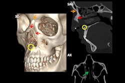
 A 67-year-old patient was brought to the emergency department after severe trauma. The CT scan of the encephalon shows a left occipital fracture with extension to the petrous portion of the ipsilateral temporalis (longitudinal type). CT angiography of the cerebral arteries reveals early repletion of the arterial phase of the left superior ophthalmic vein, as well as several hyperdense foci in the left cavernous sinus (circle), indicative of a carotid-cavernous fistula. All figures courtesy of Dr. César Antonio López López et al and presented at ECR 2025.
The orbit is delimited by four bony walls: the superior, medial, inferior, and lateral, consisting of a total of eight bones. They are classified according to the involvement of the orbital rim. In the absence of involvement of the orbital rim, they are classified by the displacement of the bony fragments outwards or into the orbit (blowout and blow-in respectively).
“In these cases, it is important to describe the type of fracture, if there is herniation of the orbital fat through the fracture or if there is entrapment of the extraocular muscles, as well as to evaluate possible eye involvement (breakage, hematomas, foreign bodies ...),” noted López, adding that other post-traumatic findings to rule out include carotid-cavernous fistula.
 A 20-year-old patient who came to the emergency department following an assault. The CT scan shows a blowout fracture of the orbital floor (circle) with herniation and entrapment of the inferior rectus muscle (arrow).
Orbital melanoma may be primary, due to local extension from the uvea, conjunctiva, or eyelid, or due to metastatic disease. Primary melanomas are extremely rare, accounting for less than 1% of orbital neoplasms, as opposed to uveal melanomas, which represent the most frequent primary intraocular tumor and account for up to 20% of secondary/metastatic tumors of the orbit.
CT can show masses with increased attenuation and enhancement after contrast administration, according to the researchers. Among the MRI findings are the following:
- On T1-weighted imaging, hyperintense due to previous hemorrhages and melanin content (after gadolinium administration, it shows enhancement)
- On T2-weighted imaging, hypointense
- On susceptibility-weighted imaging/T2*, susceptibility artifacts due to previous hemorrhages
Ocular trauma is the main cause of visual acuity loss in younger patients. One of its possible outcomes is globe rupture, an ophthalmological emergency. In blunt trauma cases, ruptures are most common immediately following insertion of the rectus muscles, they continued.
Among the different findings identified on CT are loss of the rounded morphology of the eyeball (“flat tire sign”), presence of foreign bodies or intraocular gas, thickening of the posterior sclera, and blurring of the eyeball margins.
“Orbital masses have a wide differential, and a correct description of the tumor is essential to narrow it down to the most probable pathologies,” the authors stated. “They can be classified into three main compartments: intraconal, extraconal, and intraocular.”
Primary ocular lymphoma is the most common orbital tumor, accounting for up to 50% of malignant orbital lesions. It usually affects people between 50 and 70 years of age, with a higher rate among women, and up to 17% of cases are bilateral.
 An 85-year-old patient with suspected carotid cavernous fistula due to conjunctival hyperemia that evolved over nine months. The CT scan shows a 4-mm lateral nodular thickening in contact with the posterior wall of the eyeball, lateral to the optic nerve. This lesion shows enhancement after contrast administration (arrows on CT scan, and arrowhead on T1-weighted gadolinium-enhanced MRI).
CT can show a homogeneous, isodense, or slightly hyperdense mass with the extraocular musculature and discrete enhancement after contrast administration. MRI can show similar characteristics to cerebral lymphomas:
- On T1-weighted imaging, iso/hypointense with respect to the muscles and with homogeneous enhancement after gadolinium administration
- On T2-weighted imaging, iso/hyperintense with respect to the muscles
- On diffusion-weighted imaging/apparent diffusion coefficient, diffusion restriction.
Optic nerve gliomas are rare tumors that usually affect children with neurofibromatosis type 1. They are even rarer in adults, in whom they are not related to neurofibromatosis, and they are more aggressive.
Orbital meningiomas can be primary (rare; most are optic nerve sheath meningiomas, around 20%) and secondary (the most frequent; caused by extension of an intracranial meningioma into the orbit), the researchers pointed out. They are usually diagnosed in women in the fourth decade of life, although up to 25% are diagnosed in childhood, and these tend toward more aggressive behavior. In imaging, they behave similarly to intracranial ones, with two characteristic signs being the tram-track and the doughnut, observed in sagittal and coronal reconstructions, respectively.
Vascular pathology
Carotid-cavernous fistula is an abnormal communication between the carotid circulation and the cavernous sinus. It can be classified as direct (direct communication between the internal carotid and cavernous sinus) or indirect (communication through one of the branches of the carotid circulation and the cavernous sinus). Direct communication tends to be post-traumatic and of rapid evolution, tending to be found in young men. Indirect communication is usually insidious, secondary to predisposing conditions (Ehlers-Danlos Syndrome, for example), and in postmenopausal women.
“On CT we can see indirect findings such as proptosis, edema of the orbital fat, enlargement of the extraocular musculature, as well as swelling of the cavernous sinus and arterial enhancement of the ophthalmic vein and the cavernous sinus,” they stated. “It is important to suspect this pathology in certain traumas, as it is important to diagnose it because of possible complications.”
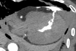
 A 94-year-old woman underwent CT following a traumatic brain injury. The CT scan shows a superoexternal orbital mass, suggestive of orbital varix.
Primary orbital varicose veins are idiopathic, while secondary varicose veins appear due to an increase in blood flow (due to arteriovenous malformations, carotid-cavernous fistulas, etc.). They are usually asymptomatic, although they may appear first in young adults with discomfort, proptosis or intermittent diplopia. In a suspicious case, a CT scan with Valsalva can be performed with contrast administration, with the consequent dilation and enhancement of the pathologic vein.
"Looking ahead, we're planning to develop follow-up work focusing on infiltrative lesions, tumors with perineural spread, and orbital involvement extending to the intracranial compartment and the pterygopalatine fossa," López, who is a second-year radiology resident, told AuntMinnieEurope.com on 10 April.
You can read the full ECR 2025 poster here. The co-authors of this exhibit were David Castanedo Vázquez, Angela Guitián Pinilla, Álvaro Sánchez Mulas, Pilar Cifrian Casuso, Elena Marín Díez, Elena Julián Gómez, Aranzazu Sánchez Gabín, and Silvia Revuelta Gómez.