Diagnostic imaging is essential for identifying colorectal cancer and determining the best therapeutic approach for the disease, according a statement issued on 31 March by the Spanish Society of Medical Radiology (SERAM).
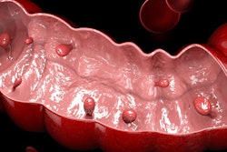
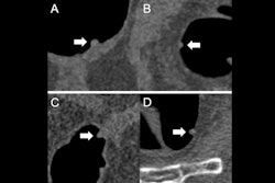
The current best practice for identifying colon cancer is colonoscopy in patients with symptoms, although screening for the asymptomatic population through fecal tests is also important, according to the society. In addition to fecal testing, virtual colonoscopy via CT imaging is well-tolerated by patients, safe, and cost-effective, SERAM said. In fact, "sensitivity for detecting colon cancer is just as good in CT as in colonoscopy (96% and 95%, respectively), and also for the detection of adenomatous polyps equal to or larger than 10 mm (94% and 98%, respectively)," SERAM noted.
Assessing results of chemotherapy or radiotherapy can be done via rectal MRI, while liver and bone lesions can be treated with percutaneous image-guided ablation procedures. And radiomics and radiogenomics can help extract and analyze quantitative information about tumors such as their heterogeneity and phenotype, according to Dr. David Leiva Pedraza, of L'Hospitalet de Llobregat in Spain.
"There is a paradigm shift in radiology, from the traditional approach of treating medical images as if they were images to a new one: correlating image characteristics with genetic, histological, clinical, prognostic and/or predictive data," Pedraza said in a SERAM statement. "[Colorectal] radiology is evolving from purely anatomical imaging techniques to quantitative, functional and molecular imaging techniques, image-based analysis of tumor heterogeneity, and the development of radiomics and radiogenomics."


