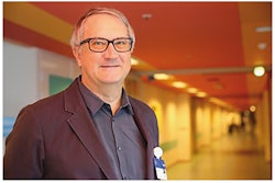
Recently, Dr. Yousef Nielsen and Prof. Henrik Thomsen wrote a thought-provoking article using a simple methodology. They looked at all the papers related to contrast media in a single journal, Acta Radiologica.1
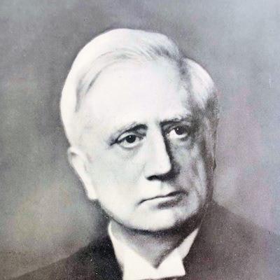
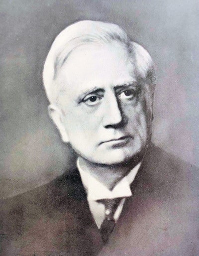 Gösta Forssell, founding editor of Acta Radiologica.
Acta Radiologica is one of the world's most significant radiological journals and was first published in 1921 under its legendary editor, Gösta Forssell (1876-1950) from Stockholm. Forssell was a remarkable man who made many original contributions to the radiological sciences and was President of the 2nd International Congress of Radiology, which was held in Stockholm in 1928.
Contrast media have been used since the earliest days of radiology, and even by 1921, the use of contrast media was well developed. Subsequent developments in medical imaging have not removed the need for them, as might have been expected. Ideally, medical imaging should be performed without the need for introducing foreign and potentially toxic material into the body.
The use of contrast media was a very hot topic when I was training in radiology in the 1980s. This was at a time when the newer nonionic media were replacing the older ionic agents. It was a fascinating time for radiology, when so many of the older ways of working were passing away and were being replaced by the developing modalities.
We had a room for renal studies -- intravenous pyelograms, or IVPs -- with two tables separated by a lead rubber screen. There was always a bowl close to hand because following the rapid injection of an ionic agent, the patients commonly vomited and experienced a strong feeling of warmth throughout their body. The newer nonionic agents came into use only slowly due to their high cost, and yet now who can remember using the conventional agents?
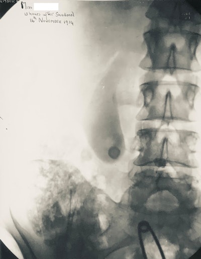 Oral cholecystogram from 1934 showing opacified gallbladder. The technique has now been replaced by ultrasound.
The history of contrast media is complex and interesting, and it was fully reviewed by Christoph de Haën in what is now the definitive history of the development of contrast media.2
The need for contrast media was well expressed by the pioneer radiologist Alfred Barclay, when he said in 1913 that x-rays penetrate all substances to a lesser or greater extent, and that the resistance that is offered to their passage being approximately in direct proportion to their specific gravity.
Barclay continued by noting that the walls of the alimentary tract do not differ from the rest of the abdominal contents in this respect, and that consequently they give no distinctive shadow on the fluorescent screen or radiograph.
Barclay clearly stated the essential problem that confronts radiologists: The density differences that are seen on the plain radiographs are those of soft tissue (which is basically water), bony and calcified structures, fatty tissues, and gas. The liver has the same density as the heart, and therefore, the two structures cannot be separated on plain films. It was only when the CT scanner was invented by Sir Godfrey Hounsfield that density differences within soft tissues could be readily appreciated, and even with CT scanning, the administration of contrast media is commonly needed.
The basis of contrast media consists in the artificial manipulation of tissue density so that specific structures are revealed, and Barclay noted that the method depends on filling the alimentary cavities with some substance that differs as widely as possible in density from that of the tissue structures, such as a bismuth salt, or by inflating them with air or other gas. Since that time all manner of solids, liquids, and gasses have been used as contrast agents.
Changing market
Nielsen and Thomsen have shown graphically the varying interest in contrast media in relation to the number of papers published. The main interest was from the 1970s to the 1990s reflecting the introduction of newer agents. There was a further peak in the 2010s reflecting recent concerns about toxicity of agents for MRI and intravascular contrast.
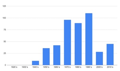 The number of publications on contrast media listed by decade.
Many agents are now obsolete. It's noteworthy that agents are developed and perfected only to become obsolete. Nielsen and Thomsen have produced graphs related to biliary and myelographic agents. It would have been most frustrating for contrast media companies to have invested so much effort in developing oral and intravenous biliary agents only to have the techniques swept away by developments in ultrasound and MRI. The authors have also put together an informative graph on acute reactions.
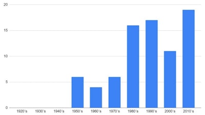 Publications on acute contrast media-related reactions by decade.
Throughout the last 100 years, there have been major developments in the field of contrast media, and the story is fascinating. The history of contrast media is brilliant!
References
- Nielsen YW, Thomsen HS. Trends in contrast media research the last 100 years. Acta Radiol. Posted online ahead of print 12 October 2021. https://www.doi.org/10.1177/02841851211051014
- Haën CD. X-Ray Contrast Agent Technology: A Revolutionary History. CRC Press.
Dr. Adrian Thomas is chairman of the International Society for the History of Radiology and honorary historian at the British Institute of Radiology.
The comments and observations expressed herein do not necessarily reflect the opinions of AuntMinnieEurope.com, nor should they be construed as an endorsement or admonishment of any particular vendor, analyst, industry consultant, or consulting group.
