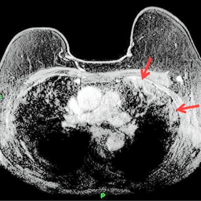
A 68-year-old English woman was treated for a likely case of COVID-19 after a radiologist spotted unusual pulmonary findings on an MRI scan for breast cancer staging. The team behind the case published a report in the September print issue of Radiology Case Reports.
The woman was asymptomatic for COVID-19 at the time of the MRI, and she only reported feeling mild upper respiratory symptoms two weeks prior to the scan. But the radiologist interpreting her contrast-enhanced MR images found signs of subpleural consolations and a high T2 signal, suggesting she was infected with the novel coronavirus.
"Given the predilection of COVID-19 for affecting the respiratory system, any imaging which encompasses all or part of the lungs could display incidental changes of the infection," wrote the authors, led by Dr. Adam Brown from Royal Free Hospital in London (Radiology Case Reports, September 2020, Vol. 15:9, pp. 1629-1632).
The woman was diagnosed with invasive lobular breast carcinoma after an abnormal mammogram result. She had been referred to the Royal Free Hospital radiology department for local staging, which showed unifocal carcinoma in the patient's upper, outer quadrant of her left breast.
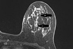
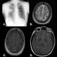
But the breast cancer wasn't the patient's only notable MRI finding. The reading radiologist also spotted incidental findings in the pulmonary space on the patient's MRI, including abnormal subpleural high T2 signal intensity in the lung periphery and enhancement in subpleural regions on postcontrast, fat-saturated T1-weighted images.
"Given the high prevalence of COVID-19 infection in the local community at the time, concern was raised that the incidental lung findings may be secondary to positive COVID-19 status," the authors wrote.
The radiology department contacted the patient, urging her to seek follow-up care at the hospital. She underwent chest radiography, which showed peripheral patchy airspace consolidation -- a finding common with COVID-19. At the time, the patient was asymptomatic, and she and her household members were instructed to self-isolate according to federal guidelines.
One day later, the patient experienced increased breathlessness and returned to the emergency department. A CT scan revealed bilateral subpleural ground-glass opacities with consolidation and multiple right lower lobe segmental acute pulmonary emboli, helping to strengthen the likelihood of COVID-19 infection.
The hospital staff treated the patient for COVID-19, according to senior study author Dr. Anmol Malhotra, consultant radiologist and breast imaging lead at the Royal Free Hospital. Malhotra noted that the patient also developed an incidental pulmonary embolism secondary to COVID-19, but she recovered and was discharged.
"This represents the first case of COVID-19 identified on breast MR imaging that the authors have seen and highlights the importance of prompt identification and flagging of incidental pulmonary findings to minimize further transmission of the virus in asymptomatic carriers," the team wrote.
While COVID-19 findings are well-documented for CT and chest radiography, the disease's presentation is not as well understood on MRI. Other reports have said COVID-19 can present similarly to other forms of pneumonia, including nonspecific high T2 signal intensity with enhancement on postcontrast T1 imaging.
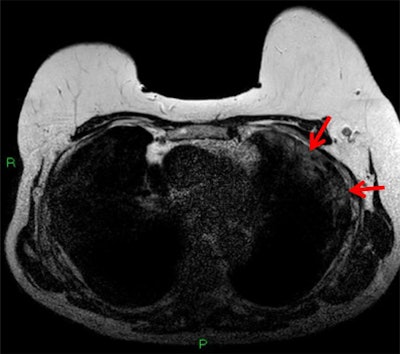 T2-weighted MRI depicting subpleural high T2 signal intensity (red arrows). All images courtesy of Dr. Anmol Malhotra.
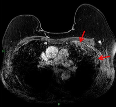 T1-weighted postcontrast fat-saturated MRI depicting subpleural consolidation enhancement (red arrows).
The case also highlights the importance of looking for incidental findings on images that at least partially visualize the lungs. This not only includes breast MRI but also shoulder trauma imaging and some abdominal CT and MRI, the authors noted.
"Identification of these findings in asymptomatic patients allows appropriate isolation measures of patient and household contacts to be commenced to minimize further spread of the virus and also allows safety net advice to be given to make the patient aware of any possible future deterioration," Anmol and colleagues concluded. "Training of radiographers to identify suspicious lung changes at the time of image acquisition can allow for appropriate cleaning of equipment and surroundings to minimize risk of infection to subsequent patients."

