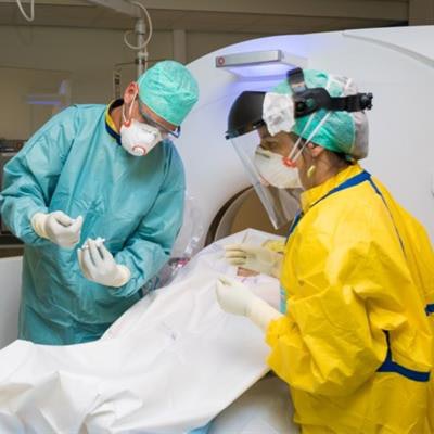
Researchers from Belgium are using CT scans to help perform minimally invasive autopsies on patients who died after being diagnosed with COVID-19.
The scans will help researchers from the Jessa Hospital in Hasselt to uncover the physical mechanisms that led to fatal COVID-19 cases for 20 patients, according to a news report posted on 8 July by the Brussels Times.
The autopsies include a comprehensive CT scan of major organs, starting with the patients' heads and ending just below their pelvis. Radiologist Dr. Jan Vanrusselt also takes needle biopsies from the lungs and other organs.
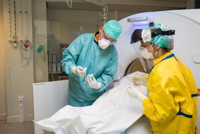 Radiologist Dr. Jan Vanrusselt takes a biopsy of a deceased COVID-19 patient. Courtesy of hbvl.be.
"First, there is the safety aspect and the reduced risk of contamination of employees," Prof. Janneke Cox, an infectious disease specialist and co-lead researcher of the project, said. "In addition, surviving relatives are also more ready to cooperate on a minimally invasive autopsy because there are hardly any injuries to the body."
More than 70% of next of kin eventually agree to such an autopsy, and relatives often go along with the idea because they feel that way they can contribute to understanding COVID-19 better, she added.
Patients with either severe acute respiratory syndrome coronavirus 2 (SARS-CoV-2) polymerase chain reaction (PCR) positivity or radiologically confirmed COVID-19 who died during hospitalization were included in the study after oral informed consent was obtained from the legal representative, Vanrusselt explained. A whole body CT-scan was performed, followed by CT-guided needle biopsies. Four sterile lung biopsies were taken for microbiology examination and at least two biopsies from heart, lungs, liver, spleen, kidneys, and abdominal fat for histological examination. Additional samples were taken when indicated.
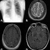
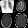
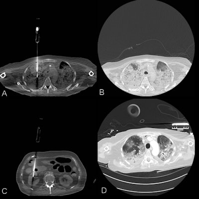 A series of annotated CT images of COVID-19 patients. A: Postmortem lung biopsy, same region as premortem CT image. B: Postmortem lung window, same region as premortem CT image. C: Postmortem renal biopsy. D: Premortem CT, 24 hours before biopsies. Courtesy of Dr. Jan Vanrusselt.
Two clinicians, two radiologists, two microbiologists, and two pathologists independently assessed the clinical file, the postmortem CT scan, microbiology findings, and histology slides respectively. The clinical cause of death and contributing diagnoses were abstracted from the discharge letter. During a multidisciplinary meeting, the multiple causes of death and contributing diagnoses were formulated and compared to the certificate of cause of death and contributing diagnoses.
Researchers at Hasselt, Ghent, and Antwerp universities then analyzed the extracted tissues to study the immunological body response and effects on tissues. The team hopes to publish their results this summer.
"Using the results of the autopsies we can find out exactly what damage the virus caused, as well as which processes are important and which are not," Cox said.
Vanrusselt said he is not aware of any other groups performing minimally invasive autopsies on COVID-19 patients.
