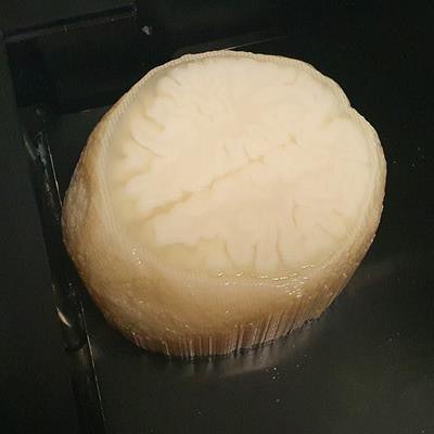
Researchers from the U.K. and Sweden have created a realistic 3D-printed brain based on MR images. The model is ideal for refining brain CT segmentation workflows, which, in turn, may improve the evaluation of neurological disorders on CT.
The group, led by Robin Holmes, PhD, of University Hospitals Bristol National Health Service (NHS) Foundation Trust, found that the 3D-printed brain phantom replicated the anatomical structure of the brain and matched attenuation properties of the originating MRI data. In addition, the team confirmed the viability of using the phantom to test the accuracy of CT segmentation workflows.
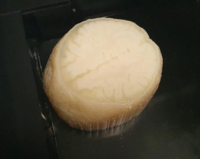 3D-printed brain model based on MR images. Image courtesy of Robin Holmes, PhD.
"This [3D-printed brain] phantom has immediate applications in dose versus image quality optimization for visual image interpretation and the selection of minimum acceptable segmentation accuracy for existing and proposed clinical CT segmentation workflows," the authors wrote in an article published online on 11 March in Medical Physics.
Brain CT segmentation
CT remains a first-line neuroimaging technique for evaluating patients suspected of having neurological conditions such as dementia. Through visual inspection, physicians are able to identify abnormalities such as cerebral atrophy and ventricular enlargement on brain CT scans.
In recent years, clinicians have sought to improve upon visual inspection by segmenting imaging data into distinct types of tissue, from soft tissue to bone to brain matter. The development of automated techniques for MRI segmentation has allowed researchers to uncover various potential benefits of using MRI segmentation for examining a wide range of neurodegenerative diseases. Yet special hardware requirements, long processing times, and strict acquisition requirements have limited the technique's clinical application.
CT segmentation offers an alternative to MRI segmentation that may be more clinically feasible due to the modality's lower cost, faster acquisition times, higher spatial resolution, and greater availability, Holmes and colleagues noted.
In the current study, the researchers set out to explore the feasibility of using CT segmentation to help examine neurological conditions, with the hope that it might reduce working time for radiologists and radiographers as well as improve diagnostic accuracy.
To that end, they turned to 3D printing technology to create a realistic anthropomorphic phantom of the brain they subsequently used to assess various CT segmentation techniques.
"Properly developed, this type of phantom will allow the optimization and validation of CT segmentation across different scanners and disease states. ... Using different acquisition parameters will enable the validation of the entire processing chain in the proposed clinical implementation of CT [segmentation]," the authors wrote.
3D-printed brain phantom
The researchers began by obtaining the brain MR images of 10 individuals between the ages of 56 and 68 from the Alzheimer's Disease Neuroimaging Initiative database. They used open-source computer software to segment the MR images and create separate stereolithography (STL) files for different types of tissue, including white matter, gray matter, and cerebrospinal fluid.
After processing the images, they input the files into a 3D printer (Objet, Stratasys) and generated a 3D-printed brain using combinations of resin-like and soft, flexible materials to represent the different types of tissue. They also made a skull-like structure out of plaster with which to surround the 3D-printed brain.
The researchers scanned the 3D-printed brain with multiple CT scanners to capture its anatomical and attenuation properties. They also applied various segmentation techniques to the CT data to assess the viability of using the phantom for CT segmentation.
Overall, they discovered that the 3D-printed brain was an accurate reflection of the source MRI data and a faithful reproduction of brain tissue and anatomy. For example, the Hounsfield unit (HU) values of gray matter ranged from 32.9 HU to 35.8 HU for the 3D-printed brain, compared with 29.9 HU to 34.18 HU reported in clinical studies. And the CT segmentations were offset from the original data by only 1 mm along the hippocampal axis and 2 mm laterally.
The segmented images, acquired using different CT scanners and different segmentation methods, also showed "excellent agreement" with the original data, with a peak Dice similarity coefficient of approximately 0.8 for gray matter.
It cost the group approximately 900 pounds (around 980 euros) to create the 3D-printed brain and took roughly two hours to generate the STL files for 3D printing.
"3D printing can be used to create a highly realistic -- in terms of both physical characteristics and anatomical accuracy -- physical CT phantom of the human brain," the authors wrote. "As the precise internal structure of the phantom is known, it was possible to demonstrate the similarity of the two [CT] scanners and the improved performance of one of the segmentation methods compared to the other."
