Patient-specific 3D-printed heart models based on cardiac MRI and CT scans are dimensionally accurate and also offer clinical value in the management of congenital heart disease, according to a new analysis published online by the Journal of Clinical Medicine.
Recently, numerous medical institutions have been exploring the potential of 3D printing to improve upon existing methods for visualizing complex congenital heart disease, which has traditionally involved looking at images on 2D flat screens, noted senior author Zhonghua Sun, PhD, and colleagues from Curtin University in Perth, Australia.
"Most of the published studies that investigated the application of 3D-printed heart models are isolated case reports or case series that are largely anecdotal. ... It remains unclear whether 3D printing can significantly improve on how the disease is managed in current clinical practice," they wrote.
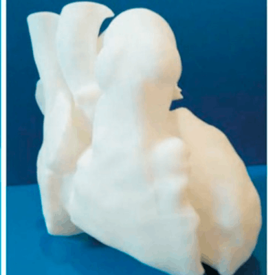
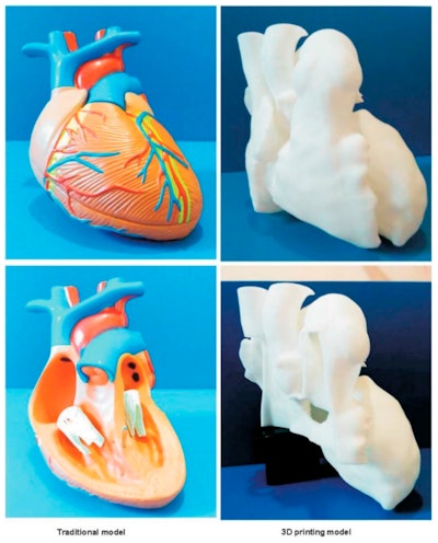 Generic heart model (left). Individually tailored 3D-printed model of a patient with congenital heart disease (right). Image courtesy of Lau et al. Licensed under CC BY-NC 4.0.
The researchers looked closely at the results from all peer-reviewed congenital heart disease studies using 3D printing that were available in several publications databases, including PubMed and Medline, as of April 2019.
In all, 24 studies met the inclusion criteria for their systematic review, and seven were eligible for meta-analysis. The predominant imaging modality used to create the 3D-printed models was CT angiography, followed by cardiac MRI and echocardiography (J Clin Med, 18 September 2019).
The group found a strong correlation between the dimension of the 3D-printed heart models and the patient MRI and CT scans (r > 0.98). Although the models did slightly underestimate the size of patient anatomy, with an average deviation of 0.04 mm compared with the medical images, this difference falls well within the acceptable range of deviation in daily clinical routine, according to the researchers.
Sun and colleagues also identified four main clinical applications of 3D printing in the context of congenital heart disease, including the following:
- Preoperative planning. By far the most common application, using 3D-printed models to plan surgery helped surgeons appreciate the complexity of cardiac anatomy and visualize abnormalities that they could not clearly identify on cardiac CT and MRI alone. The models also helped surgeons decide on the best surgical approach and even altered the surgical decision in nearly half of the cases for one study.
- Medical education. Collectively, the studies showed that using 3D-printed anatomical models for medical education did not lead to any statistically significant improvements in knowledge-based test scores. Yet one study did show statistically significant improvements in test scores when using 3D-printed models to educate participants about complex (but not simple) congenital heart disease, suggesting that the models could have a role in teaching complex heart pathology. Furthermore, all of the studies' investigators reported improvements in subjective evaluation scores and satisfactory level among participants and clinicians.
- Patient consultation. Questionnaires from the studies indicated that 3D-printed models were useful in enhancing communication between providers and patients' parents, with some physicians claiming the models resulted in a better interaction with parents, compared with conventional images alone. The average duration of patients' consults was also several minutes longer when 3D-printed models were involved. Rather than replace the traditional approach of communicating with patients using 2D medical images, the 3D-printed models seem best suited as a complementary tool to standard images for patient consults, the authors noted.
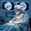
- Intraoperative orientation. In several studies, surgeons relied on a 3D-printed heart model to orient themselves to patient anatomy during surgery, reducing the operating time.
Among the various applications of 3D printing in congenital heart disease, the technique seems to play the most important role in facilitating preoperative planning for complex cases, the authors explained. It may still be too early to determine the clinical value of 3D printing for medical education as well as the extent to which it might help improve patient outcomes, but this early research demonstrates the technology has promise.
"Last but not least, a comprehensive cost-benefit analysis for implementation of 3D printing technology in cardiovascular surgeries lacks in the current literature and this needs to be addressed by future studies," the authors concluded.

