Spanish researchers testing a dedicated breast PET scanner found that it had a high false-negative rate for detecting in situ carcinomas in women with BI-RADS 4 lesions. The finding raises questions about the suitability of the system for working up suspicious breast lesions, according to a 3 October study in the European Journal of Radiology.
In a group of 50 patients with mammograms classified as BI-RADS 4, dedicated breast PET missed nine of 18 cancers. In addition, dedicated breast PET recorded increased metabolic activity in 10 benign lesions.
"Our analysis does not allow the recommendation of dedicated breast PET for diagnosis of malignancy in BI-RADS 4 mammographic or ultrasound abnormalities, given the high rate of false-negative results regarding in situ neoplasms," wrote lead author Dr. Lucía Graña-López from Hospital Lucus Augusti Lugo and colleagues.
Molecular breast imaging has come to the forefront in recent years as a tool that could help with the detection of breast cancer, especially among patients classified as BI-RADS 4 or greater. While mammography and ultrasound are generally the modalities of choice for workup, they also can produce a high number of false positives, as does breast MRI.
Could molecular imaging offer an alternative? The group from Hospital Lucus Augusti Lugo decided to investigate the potential of a dedicated PET scanner, (Mammi, OncoVision), that is available in Europe.
In this study, the researchers enrolled 50 consecutive women (median age 48 years; range, 21-75 years) who presented with a total of 60 BI-RADS 4 lesions. All patients underwent bilateral full-field mammography followed by sonographic exams.
In addition, before biopsy, subjects also underwent 1.5-tesla breast MRI scans, with the dedicated breast PET exam following. The researchers then fused MRI and dedicated breast PET images to better locate corresponding lesions and to avoid misinterpretation of dedicated breast PET results.
All findings were compared with histological results, which confirmed 42 benign lesions (70%) and 18 malignancies (30%). Of those malignancies, seven lesions (39%) were in situ and 11 (61%) were invasive carcinomas.
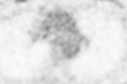
Dedicated breast PET discovered 19 lesions; 10 were benign and nine were malignant. However, the modality missed a significant number of carcinomas in situ and approximately one-fourth of invasive cancers. In other words, there were nine (50%) false-negative results in the sample.
| Performance of dedicated breast PET in BI-RADS 4 lesions |
|
| Sensitivity | 50% |
| Specificity | 76% |
| Percent of invasive carcinomas detected | 73% |
| Percent of in situ carcinomas detected | 14% |
"Two invasive carcinomas were located less than 1 cm from the pectoral muscle, which can explain that they were missed by dedicated breast PET, because they were outside the field-of-view," the authors noted.
Graña-López and colleagues recommended additional research with larger cohorts to better evaluate the role of dedicated breast PET with BI-RADS 4 lesions and "as a complementary tool to conventional breast imaging techniques."

