Researchers from the Netherlands have developed a method to quantify wrist motion using 4D CT, according to a Thursday presentation at the Computer Assisted Radiology and Surgery (CARS) 2018 international congress in Berlin. The technique may help improve bone displacement and fracture healing evaluation.
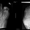
The wrist is composed of an intricate collection of bones with different shapes that can superimpose on each other and make it difficult for clinicians to visualize using conventional radiography. Other imaging modalities including MRI and conebeam CT have shown promise toward improving visualization in the area, especially when supplemented with volume rendering or other 3D imaging techniques.
Taking these enhancements one step farther, a team of investigators, led by Dr. Geert Streekstra from Amsterdam University Medical Center, found a way to view wrist movement over time on CT scans.
For this process, the patient table stays in a fixed position inside the CT scanner while the patient slowly moves the wrist and the gantry rotates, acquiring one scan per revolution, presenter Dr. Iwan Dobbe, PhD, told AuntMinnieEurope.com. The resulting CT scans are then converted into a series of 3D models that collectively allow viewers to see the wrist in motion, or 4D CT.
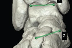
The group quantitatively analyzed wrist motion by using proprietary software to segment relevant bone structures from the initial CT scan and to register each bone to every frame in the 4D CT model.
"The technique allows us to show the bones in each 4D frame and to quantify bone displacements throughout the 4D scan," he said. "This helps us in judging if bone displacements are normal or pathological."
In addition, they distinguished actual wrist motion from methodological errors with 4D CT by altering the wrist's speed and axis of motion as well as the gantry's revolution speed. They observed the highest accuracy and least motion blur in bone displacements when the wrist was moving slowly and the CT scanners had a high gantry revolution speed.
Recently, the group applied this technique to clinical scenarios involving wrist fractures -- particularly for the diagnosis of scaphoid fractures.
"We are already involved in clinical 4D CT studies to better diagnose if a fractured scaphoid bone resulted in an unstable nonunion with segments that are loose," Dobbe said. "We will also utilize the method to copy the kinematics of a healthy arm for use in an injured arm that needs a patient-specific kinematic implant."


