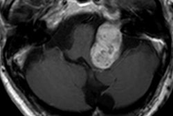
In the routine follow-up of patients with intracranial meningioma, unenhanced MRI scans are as effective in evaluating the disease as imaging with gadolinium contrast, according to a study from Turkey presented at ECR 2018 in Vienna.
In comparing precontrast and postcontrast MR images, the researchers discovered the size of meningiomas in both datasets showed tumor growth. Given some concern over the use of gadolinium-based contrast agents (GBCAs), the option not to use the agents at all could put clinicians and patients at ease and produce similar results, noted lead author Dr. Feride Kural Rahatli from Başkent University in Ankara.
"Although the clinical effect of gadolinium deposition is unknown, in order to avoid gadolinium deposition in patients with meningiomas, they can be followed up with unenhanced brain MRI scans," she said.
Brain MRI with GBCAs is routinely used in the follow-up of meningiomas with linear agents being widely used, Rahatli told ECR attendees, and hyperintensity on axial T1-weighted images has been shown with serial brain MRI using linear GBCAs. Still, clinicians and patients must weigh the risk against the benefit when it comes to using a GBCA for an MRI scan, she explained. Patients with adequate renal function are considered not at risk for an adverse reaction if they can evacuate the contrast from their bodies, so perhaps the best solution is to avoid GBCAs whenever possible.
"We aim to show that unenhanced brain MRI can be used in routine follow-up of patients with intracranial meningioma in order to avoid gadolinium deposition in the brain by measuring the size of a meningioma on precontrast T2-weighted MR images," Rahatli said.
Patient details
This retrospective study enrolled 29 patients (age range of 26 to 86 years old; median age of 61) between November 2010 and October 2016. All the subjects had estimated glomerular filtration rate (eGFR) greater than 60 mL/minute. Patients with an eGFR below 30 mL/min were not given gadolinium-based contrast.
The GBCA-enhanced brain MRI scans were performed on a 1.5-Tesla device (Avanto, Siemens Healthineers). Administration of gadoversetamide (Optimark, Guerbet) was based on 0.001 mmol or 0.2 mL/kg. The protocol included axial T1- and axial T2-weighted imaging, as well as axial postcontrast T1-weighted imaging.
The dimensions of the 30 discovered meningiomas were measured from the anteroposterior and mediolateral based on the first and last MRI scans on both axial precontrast T2-weighted images and axial postcontrast T1-weighted images.
To determine gadolinium deposition, the researchers drew regions-of-interest (ROI) on the axial precontrast T1-weighted images from the dentate nucleus, pons, and globus pallidus. Signal intensity ratios were calculated for dentate nucleus-to-pons, dentate nucleus-to-cerebellum white matter, and the globus pallidus-to-thalamus.
In two patients, globus pallidus-to-thalamus signal intensity ratios were measured from the left side because of chronic encephalomalacia in the right basal ganglia. The other 29 measurements were all taken from the right side of the brain. The meningioma ranged in size from 6 x 5 mm to 45 x 40 mm. A total of 22 meningiomas were supratentorial, and eight meningiomas were infratentorial.
Signal intensities
In comparing MRI scans from baseline to follow-up, six meningiomas in five patients increased in size based on measurements from both axial precontrast T2-weighted images and the axial postcontrast T1-weighted images. The mean increase in anteroposterior length was 7 mm (range, 5 to 13 mm), while the mean increase in mediolateral length was 6 mm (range, 5 to 7 mm).
Despite the size increase, none of the patients showed clinical symptoms of the disease after the brain MRI scans.
In reviewing the signal intensity ratios, the researchers did see a statistically significant increase (p < 0.05) between the first and second MRI scans in all regions.
| Signal intensity ratios of MRI scans | |||
| First MRI | Second MRI | P value | |
| DN/pons ratio | 1.055 ± 0.38 | 1.149 +/1 0.86 | < 0.05* |
| DN/cerebellum | 1.068 ±0.42 | 1.156 ± 0.78 | < 0.05* |
| GP/thalamus | 1.077 +/0.24 | 1.136 +/ 0.58 | < 0.05* |
Given the similar sizes and equal growth of meningiomas in postcontrast axial T1-weighted images and precontrast axial T2-weighted images, and increase in signal intensity from the administration of the GBCA in patients with normal renal function, Rahatli and colleagues concluded that gadolinium contrast and subsequent deposition could be avoided in patients with meningioma using unenhanced brain MRI for follow-up scans.


