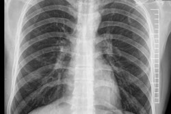
Radiologists who make errors are not always negligent, sloppy, or careless, but a greater emphasis on training and competency as well as improvements in systems used for reporting and communication of images can reduce the number of discrepancies and clinical incidents, U.K. researchers have found.
Radiology involves decision-making under conditions of uncertainty, and radiologists cannot always produce infallible interpretations, noted Dr. Amanda Jewison, a radiologist at Brighton and Sussex University Hospitals Trust, U.K., and colleagues in an e-poster presentation at RSNA 2017.
"Errors in radiology, as in life, are inevitable," they stated. "It is not a binary process. The correct answer is not always 'normal' or 'abnormal;' 'cancer' or 'not cancer.' The final radiology report is influenced by many variables."
The term "error" is often an unsuitable term to use when something goes wrong, because error implies a mistake -- i.e., an incorrect interpretation of an imaging study. Therefore, it may be more appropriate to concentrate on a "discrepancy" between a report and the review of a particular outcome, added Jewison and colleagues, who presented imaging cases taken from a cross-departmental clinical governance round in which highlighted errors and discrepancies documented by pulmonologists were discussed with the radiology department in a process of shared learning.
Perceptual errors in chest imaging
A perceptual error is deemed to have occurred when an abnormality is retrospectively determined to have been present on a diagnostic image, but was not seen by the interpreting radiologist at the time of primary interpretation. These occur during the initial detection phase of image interpretation, they explained.
"For a report to be erroneous, it follows that a correct report must be possible, i.e., it fails to reach the same conclusion that would be reached by a group of one's peers. Errors can only arise where the correct interpretation is not in dispute," they continued.
Previous studies have shown that experienced radiologists failed to note important findings on 30% of chest radiographs that were positive for disease. Also, between 70% and 90% of lung cancers are visible in retrospect on previous chest x-rays (CXR) reported as normal, and 19% of lung cancers presenting as a nodular lesion are missed.
A 3% to 5% real-time error rate in radiologists' day-to-day practice is thought to be the average, according to Jewison and colleagues. Prolonged attention on a specific CXR area ("visual-dwell") tends to increase the number of false positives and false negatives, and reducing the viewing time to four seconds leads to a higher miss rate in most cases.
"The cause of perceptual errors is poorly understood but is more frequent when the target lesion is poorly conspicuous, there is reader fatigue, a rapid pace of interpretation, distractions (e.g., phone/email) and "satisfaction of search,' " the authors noted.
Most common locations
The most common locations for missed pathology on chest radiographs are the lung apices, the hilar regions, paratracheal stripe, and the retrocardiac region and area posterior to the diaphragms. In the U.K., lateral radiographs are not performed routinely, increasing the need for robust review areas, and these can be incorporated into a checklist as part of a structured or template report, they wrote.
Superimposition of lung nodules and osseous structures has been shown to reduce sensitivity in lung nodule detection on CXR. Improvement in detection and characterization has been demonstrated with dual-energy subtraction radiography and digital tomosynthesis techniques.
Growing evidence suggests that dual-energy CXR improves confidence and accuracy in the diagnosis of pulmonary nodules. Research suggests improvement can occur in sensitivity for detection of previously missed small lung cancers from 40% to 59%, with no increase in false positives. Increasing the speed of CXR reading from 13 to 10 seconds for nodule detection can lead to greater confidence in a negative read.
The limitations of CXR for lung nodule detection are well known, the authors pointed out. Prospective data from screening trials have shown no survival benefit from screening CXR in at-risk patients compared with standard care, while national lung screening trials have led to a 20% mortality reduction between CT- and CXR-screened participants.
Medicolegal perspective
Judging perceptual errors in law has proved complex, they added. The legal standard for negligence involves proving a breach in the usual standard of care, i.e., the same degree of care, knowledge, and skill as an average physician. Radiological errors are difficult to assess objectively due to the concept of hindsight bias, particularly when people with knowledge of the outcome of an event believe falsely they would have predicted it. In so-called creeping determinism, outcome information is integrated into someone's knowledge of events preceding that outcome.
Most legal jurisdictions have established the doctor cannot be perfect. In fact in cases relating to radiology, judges should instruct juries that, as Caldwell pointed out in 2007 in Ann Health Law:
[T]here is an absolutely unavoidable "human factor" at work in the review of films; some abnormalities may be missed, even the obvious ones; the mere fact that a radiologist misses an abnormality on a radiograph does not mean that he or she has committed malpractice; and not all radiographic misses are excusable. Therefore, the focus of attention should be on issues such as proof of competence, habits of practice, and use of proper techniques.
Cognitive and system errors explained
Cognitive or interpretive errors occur when an abnormality is identified but its importance is incorrectly interpreted. This type of error may be secondary to a lack of knowledge, a cognitive bias or misleading clinical information, or it may be a result of a radiologist inadvertently propagating an error made by a colleague in a previous radiology report, according to Jewison and colleagues.
Examples include complacency (a finding is appreciated but attributed to the wrong cause); faulty reasoning (a finding was appreciated and interpreted as abnormal, but attributed to the wrong cause); lack of knowledge or subspecialist training on the part of the viewer; under reading or satisfaction of search, where detection of one abnormality on a radiographic study results in a premature termination of the search, allowing for the possibility of missing other, related or unrelated abnormalities; poor communication (i.e., failure to communicate critical, urgent, or unsuspected findings to the referrer); the lesion was identified and interpreted correctly, but the message failed to reach the relevant clinician; and complications, most frequently during invasive procedures.
An example of an interpretative error is a CXR in a patient with chronic cough reported as demonstrating reticular opacities consistent with interstitial lung disease, they wrote. On subsequent review of the CXR and a CT scan, the diagnosis of bronchiectasis is made.
Errors in interpretation are multifactorial and include lack of relevant clinical information, inadequate training or subspecialization of the radiologist for a particular examination, and legitimate differences of opinion between experts, the authors elaborated.
Examples of system errors are excessive workload; degradation of lung cancer detection with decreased viewing time, and increased error rates in abdominal CT reporting when the radiologist reports more than 20 studies per day; inadequacy of clinical information; clinical diagnosis has been shown to change in 50% of cases following communication between clinician and radiologist, with a change of treatment in 60% of cases discussed; knowledge of pertinent clinical history has been shown to increase the accuracy of CXR interpretations from 38% to 84%; inappropriate expectations; capability of a particular radiologic technique may not be suitable for the question being posed; unavailability of previous studies or reports for comparison; over-reliance on locum radiologists; inexperience of staff; and inadequate equipment.
Failure to effectively communicate results in a timely manner is a potential source of error and suboptimal patient management, but patient harm may be prevented by direct communication of urgent findings, they continued.
American College of Radiology (ACR) guidelines advise radiologists to speak directly with the referring physician and document the communication in the radiology report. When a direct conversation with the referring clinician is not possible or practical, the U.K. Royal College of Radiologists (RCR) advises the report is red-flagged and urgently faxed/emailed to the referring team with appropriate checks in place to ensure this is acknowledged (e.g., OrderComms), Jewison and colleagues concluded.


