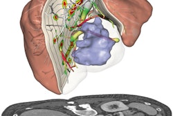
Artificial intelligence (AI) can accurately segment the liver on contrast-enhanced MRI examinations, potentially yielding significant time savings for radiologists without affecting the accuracy of volumetry results, according to researchers from the University Hospital Regensburg in Germany.
Using a small training and validation dataset, a team led by Dr. Niklas Verloh found that a convolutional neural network (CNN) could provide a high level of segmentation performance. What's more, volumetry results can be provided in just seconds, compared with approximately 10 minutes for expert human readers.
Consequently, the neural network could help radiologists by offering a fully automatic liver segmentation scheme for contrast-enhanced MRI images, according to Verloh. He presented the research, which received a student travel stipend award, at the RSNA 2017 meeting in Chicago.
A crucial task
Liver volumetry is a crucial task. It's needed during treatment planning of patients with a chronic liver disease, to determine patient-specific liver anatomy for patients undergoing liver surgery, and to calculate the hepatic functional reserve. However, the preferred segmentation method -- freehand drawing of the liver by radiologists -- is time-consuming and prone to human errors such as overestimation due to the inclusion of vessels or liver tumors, Verloh said.
After reviewing the literature, the researchers found liver segmentation remains an open challenge for computer-based approaches. It's a complicated task due to the high variability of the liver size and shape between patients, low contrast between adjacent tissues or organs, and the presence of pathologies such as a tumor or cirrhosis, he said.
There are some semiautomated and fully automated software applications available for estimating liver volume, but those are mostly for use with CT scans, Verloh said. However, MRI is increasingly utilized for liver resection and transplant planning.
"We thought, 'Why isn't there one for liver MRI?' " Verloh said.
A reliable, automated method
As a result, the researchers sought to develop a fully automated and reliable liver volumetry method in contrast-enhanced MRI based on deep-learning algorithms. First, they gathered 48 datasets of contrast-enhanced liver MR images. All studies were performed on a Magnetom Skyra 3-tesla MRI scanner (Siemens Healthineers) using T1-volumetric interpolated breath-hold examination (VIBE) sequences with fat suppression acquired 20 minutes after contrast injection of Primovist (Bayer HealthCare Pharmaceuticals). The datasets were then split into 44 training cases and four validation cases.
For the purposes of the study, the manual segmentations initially performed by a resident physician with five years of hepatobiliary imaging experience were used as the gold standard. In addition, nine cases were also cross-segmented by an additional reader with five years of experience to determine the experts' segmentation overlap, Dice index, and intraclass correlation coefficient. The researchers used a neural network topology based on the V-Net CNN, which is derived from the U-Net CNN architecture.
In testing, the network was able to predict the segmentation of a 64-slice MRI scan in approximately four seconds. In comparison, manual segmentation took approximately 10 ± 2 minutes per case, he said. In addition to being significantly faster than the human experts, the algorithm yielded a higher concordance with the ground truth.
| Comparison of epxerts and neural networks | ||
| Expert 1 vs. Expert 2 | Neural network vs. ground truth | |
| Segmentation overlap | 90.9 ± 4.9% | 92.2 ± 0.94% |
| Dice coefficient | 95.2 ± 2.8% | 95.2 ± 2.2% |
| Intraclass correlation coefficient | 0.973 | 0.984 |
"The neural network achieves a higher concordance with the ground truth than two expert readers agree in terms of ICC and overlap," he said.
The study shows promising results with the small training and validation sample size. With the neural network, the researchers were able to provide a fully automatic segmentation scheme for MRI that was able to segment the liver parenchyma in seconds with a high intraclass correlation coefficient, according to Verloh.


