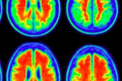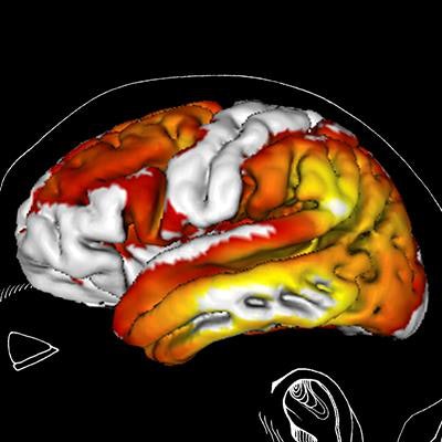
Using PET with the novel radiotracer fluorine-18-AV-1451 that binds to tau deposits, Swedish researchers have linked certain regions of the brain with the early or late onset of Alzheimer's disease and its symptoms, according to a study published in the September issue of Brain.
AV-1451 uptake in areas of tau accumulation in the neocortical regions is early indications of Alzheimer's disease, while peak AV-1451 uptake in the medial temporal lobe is associated with late-onset Alzheimer's. In addition, the early-onset group exhibited greater uptake in prefrontal, premotor, and inferior parietal cortex than late-onset subjects (Brain, September 2017, Vol. 140:9, pp. 2286-2294).
"These preliminary findings indicate that age may constitute an important contributor to Alzheimer's disease, highlighting the potential of tau PET to capture phenotypic variation across patients with Alzheimer's disease," wrote lead author Dr. Michael Schöll and colleagues from Lund University and the University of Gottenberg in Sweden.
Tau pathology
The accumulation of tau protein in the brain has been linked to neurodegenerative disorders such as dementia, Alzheimer's, and, in some cases, chronic traumatic encephalopathy. Currently, PET with AV-1451 is limited to research applications, but the imaging technique has shown early promise to evaluate neurofibrillary tau pathology in vivo.
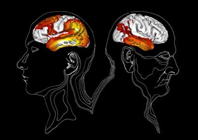 PET images show the differences in tau pathology and regions of AV-1451 uptake for early-onset Alzheimer's (left) and late-onset Alzheimer's (right). Patients with early-onset Alzheimer's (before age 65) show significantly higher uptake in lateral, temporal, parietal, frontal, and occipital lobe regions, compared with late-onset Alzheimer's (after age 65), which demonstrated the possibility to distinguish between the two conditions based on age using AV-1451-PET. Images courtesy of Drs. Annie Hallén/Michael Schöll.
One German study found a significant correlation between increased tau and decreased metabolic activity in the brain through AV-1451-PET. Such hypometabolism often is an indication of neurodegeneration. The findings also suggested that tau deposits may be more influential in causing Alzheimer's disease than amyloid accumulation.
In another previous study, German researchers conducted a multimodality investigation that used AV-1451 with PET to measure tau protein deposits; carbon-11-labeled Pittsburgh compound B to measure amyloid plaques; and FDG to measure regional neurodegeneration to explore neurodegenerative characteristics of Alzheimer's disease. The images illustrated how tau pathology may be instrumental in creating disease-modifying strategies. The work also received the 2016 Image of the Year Award from the Society of Nuclear Medicine and Molecular Imaging (SNMMI).
In the U.S., researchers at Mount Sinai in New York City used AV-1451 (also known as T807) with PET to image the brain of a living 39-year-old former professional football player who experienced 22 concussions during his career. T807-PET showed its efficacy in diagnosing concussion-related brain degeneration for the first time in a live subject. At Washington University in St. Louis, Missouri, researchers concluded that PET with AV-1451 correlated with regionally specific cortical thinning in patients with Alzheimer's disease.
Alzheimer's typically presents itself when people reach 65 years or older. At that age, patients are considered to have a late onset of the disease. However, between 5% and 10% of Alzheimer's patients show signs of the conditions long before they reach age 65. That timing is deemed as early onset.
Thus, Schöll and colleagues sought to take their AV-1451-PET research one step further and investigate how age factors into Alzheimer's and what differences there are, if any, between early-onset and late-onset patient groups.
Patient groups
Participants for this paper were recruited from the prospective and longitudinal Swedish BioFinder study, which brings together researchers, scientists, universities, and the private sector to gather information about Alzheimer's, Parkinson's diseases, and other neurodegenerative disorders. The study also develops tests for early diagnoses and effective follow-up treatments.
From BioFinder came 30 cognitively healthy elderly subjects 60 years of age and older who achieved scores of 28 points or more on the Mini-Mental State Examination (MMSE) at the study's start. An MMSE score of 24 or greater indicates normal cognition. The healthy controls also had no signs of cognitive impairment or any dementia.
The researchers also enrolled 16 patients with mild cognitive impairment due to Alzheimer's disease, early onset of the disease, or late onset of Alzheimer's. The subjects were between 60 and 80 years old and were referred to the memory clinics because of cognitive impairment, did not meet the criteria for dementia, or had an MMSE score of 24 to 30 points. They also had low cerebrospinal fluid and beta-amyloid levels.
There also was a cohort of 41 Alzheimer's patients, which include 20 subjects with early onset and 21 individuals with late onset of the disease. All 41 patients met the criteria for dementia.
Subjects underwent structural MRI on a 3-tesla scanner (Tim Trio, Siemens Healthineers) with a protocol that included T1-weighted magnetization-prepared rapid gradient echo (MPRAGE) imaging. MR images were acquired for volumetric analysis, PET image coregistration, and template normalization. PET scans (GE Discovery 690, GE Healthcare) were performed 80 to 120 minutes after a bolus injection of 370 MBq of AV-1451.
AV-1451 uptake
Through the AV-1451-PET images, the researchers discovered unique uptake patterns in both early- and late-onset Alzheimer's patients. In a comparison with healthy controls, the early-onset disease group showed greater uptake in most of the temporal, lateral parietal, lateral occipital, and dorsal frontal cortical regions. Uptake among the late-onset Alzheimer's patients was greatest in the medial temporal lobe.
When the researchers matched subjects with Alzheimer's, the early-onset group had significantly more uptake in the dorsolateral prefrontal, premotor cortex, and the inferior parietal lobule (p < 0.001) than the late-onset subjects.
"The changes in the various parts of the brain that we can see in the images correspond logically to the symptoms in early onset and late onset Alzheimer's patients respectively," the authors wrote. "Our findings suggest that the inferior and medial temporal cortex may be the most optimal regions of interest to yield maximum diagnostic accuracy across the wide clinical spectrum of Alzheimer's disease."
The Swedish researchers noted several limitations with the study, including the small number of subjects in the Alzheimer's disease patient group. They added that future studies should include a larger patient cohort to determine differences at the pre-dementia stage.
In addition, because AV-1451-PET is still in its preliminary stage, the authors noted they did not obtain sufficient control data to enable proper age-matching with the respective patient groups.


