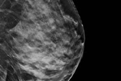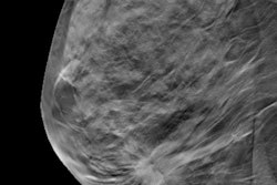
When it comes to tumor margins in specimen radiography, digital breast tomosynthesis (DBT) is king, according to a new study from Germany. The researchers found DBT tops full-field digital mammography (FFDM) in identifying the closest tumor margin and has higher sensitivity for assessing margin status.
More than 100 patients underwent breast-conserving surgery in this prospective study. After each lesion excision, specimens were marked and imaged using FFDM and DBT. The research team found that DBT is more accurate and more sensitive.
"DBT is a promising modality in performing specimen analysis," the study authors wrote (European Journal of Radiology, August 2017, Vol. 93, pp. 258-264). "It significantly improves the accuracy of specimen radiography regarding identification of the closest margin and sensitivity regarding margin status assessment compared with FFDM. This could reduce re-excision and reoperation rates."
The team was led by Dr. Heba Amer from the radiology department at Zagazig University Hospitals in Zagazig, Egypt, and the radiology clinic at Charité -- Universitätsmedizin in Berlin.
How DBT compares
The goal of any breast-conserving surgery is to achieve complete excision of the malignant lesion with adequate tumor-free resection margins because the risk of local recurrence, and poorer prognosis, is directly related to positive tumor margins, Amer and colleagues explained. Thus far, it is still difficult to achieve tumor-free resection margins during the first attempt, with re-excision rates ranging from 20% to 60%.
Specimen radiography is a technique that examines the integrity of the resection margin while the patient is still in the operating room, guiding the surgeon in either completing the operation without additional excision or extending the resection, they added.
The problem is the current techniques for performing specimen radiography using FFDM have low sensitivities compared with final histopathological margin analysis. Could DBT be the golden ticket? That's what the researchers sought to find out.
They enrolled 102 consecutive patients, each undergoing ultrasound/mammography-guided wire localization for their lesions followed by FFDM and DBT (both, Mammomat Inspiration, Siemens Healthineers). Two blinded radiologists independently analyzed the images and identified in which direction the lesion was closest to the specimen margin and measured the margin width.
The readers rated images from each modality for the presence or absence of the lesion within the specimen, lesion conspicuity, lesion type and features, lesion size, and identification of the direction in which the lesion is closest to the specimen edge. The readers' findings were corroborated by histopathological analysis.
The histopathology revealed pure invasive carcinomas in 26 cases (25%), ductal carcinoma in situ (DCIS) in 16 (15%), and a combination of invasive and in situ cancers in 60 (60%) of the specimens. In 33 cases, the margins were cancer-free and identified as clear. In 69 patients, the resection margin was positive for cancer cells.
"The high number of involved margins could be explained by the strict protocol we follow in our institution in specimen imaging (> 5 mm for DCIS, > 1 mm for invasive cancers), which is smaller than those limits applied in other studies which used 1 cm limit for both invasive cancers and DCIS," the authors wrote.
The first reader correctly identified margin direction 44% of the time using FFDM. The second reader correctly identified margin direction 36% of the time using FFDM. Using DBT, their identification was correct 68% and 69% of the time, respectively.
| FFDM vs. DBT for specimen radiography | ||
| FFDM | DBT | |
| Sensitivity for margin status | 62% | 77% |
| Specificity for margin status | 97.4% | 77% |
| Positive predictive value | 98% | 89% |
| Negative predictive value | 58% | 61% |
| Accuracy | 73% | 77% |
"The significant improvement in closest margin identification by DBT can be attributed to the better delineation of tumor margins and elimination of possible tissue overlapping over parts of the lesions," the authors wrote.
The study has some drawbacks, however, with the largest being that different pathologists performed the histopathological exams without the use of standardized procedures for correlation of radiologically and pathologically measured directions. Also, strict commitment to the marking protocol cannot be guaranteed, even if it is a standard, defined procedure.
Nonetheless, DBT is promising because in this study it significantly improved the accuracy and sensitivity in comparison with FFDM, the authors concluded.


