Radiologists at the Technical University of Munich (TUM) are using dual-energy spectral CT to improve treatment planning for pancreatic cancer patients and to gain a better idea of their prognosis. They're convinced the procedure makes it much easier to see tumor boundaries, and they hope it will provide new insights into the tissue structure and biological behavior of tumors.
Conventional clinical CT diagnosis uses 70-kiloelectron-volt (keV) x-ray tubes to generate images. It gives a very clear picture of anatomical structures, but has a limited ability to distinguish between tissues with similar x-ray properties. Dual-energy CT resolves this by using two different voltages, explained Dr. Fabian Lohöfer, a radiologist at the TUM, during the German Radiology Congress (Deutscher Röntgenkongress, RöKo 2017).
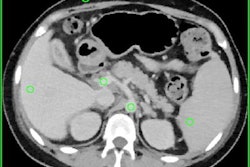
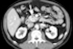
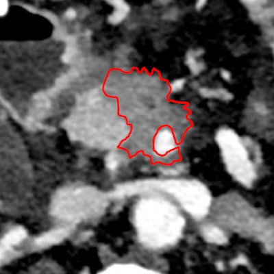
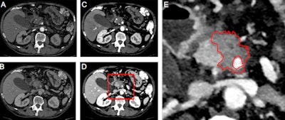 A comparison of images using conventional CT (A and B) and spectral MonoE 40-keV images (C and D) in the arterial and venous portal contrast medium phases. The images show a pancreatic carcinoma with encasement of the superior mesenteric artery. The tumor boundaries can be seen much more clearly in C and D. E shows the detail, with tumor boundary marked. All images courtesy of the German Radiological Society.
Dual-source CT is a clinically widespread dual-energy procedure that replaces a single tube with two: one of 70 and the other of 40 keV. Dual-layer spectral CT, on the other hand, uses one tube but two detectors in series.
"This makes it possible to see the whole radiation spectrum emitted by the tubes," he said.
The technology is particularly useful in pancreatic cancer, for which the prognosis is relatively poor. There are different subtypes and sensitivities to the existing standard chemotherapy, and if doctors can distinguish between these patients, they will have a clearer idea of which ones will benefit from a particular chemotherapy or operation, according to Lohöfer.
Identification of cell-rich tumors
Dual-layer spectral CT is better than conventional, contrast-medium-based CT when it comes to assessing patient prognoses, he said. "It shows the effect of the contrast agent much more clearly. We can even assess the iodine enrichment of the tissue, so we can differentiate between cell-poor tumors with a better prognosis and cell-rich ones whose outlook is poorer."
 Dr. Fabian Lohöfer.
Another advantage of dual-layer spectral CT is that it gives a clearer visual picture of tumors, with a clear distinction between tumor and healthy tissue, and of blood-vessel involvement, which is critically important in preoperative planning. It's particularly good at defining smaller tumors, he elaborated. The technology benefits not only surgeons in carrying out operations, but also physicians who want to use CT to establish whether a specific therapy will work.
Lohöfer emphasized the more accurate diagnosis offered by dual-spectral CT will benefit many patients with other cancers. "In our clinic, wherever possible, we're trying to examine all tumor patients with dual-layer technology," he said.
Computer analysis algorithms could provide even more information in future. Lohöfer believes that dual-layer spectral CT is particularly suitable for big data approaches because it provides more imaging information than conventional CT, and thus a broader basis of data for analysis.
Editor's note: This is an edited version of a translation of an article published in German online by the German Radiological Society (DRG, Deutsche Röntgengesellschaft). Translation by Syntacta Translation & Interpreting. To read the original article, click here.


