What do you really need to know about CT of peritoneal metastases? Radiologists from Barcelona, Spain, have addressed this tricky topic and collected a prestigious international prize for their efforts.
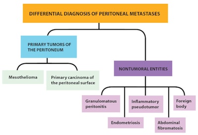
Differentiating peritoneal metastases is difficult because imaging features overlap with primary peritoneal tumors and a few nontumoral entities; often the final diagnosis must be pathological, but there are useful clues that can help to make a differential diagnosis, according to Dr. Javier Miguez Gonzalez, from the Department of Radiology at Hospital Sant Joan Despi Moises Broggi.
The peritoneal ligaments and mesenteries are double folds of peritoneum that support the intraperitoneal organs and subdivide the peritoneal cavity into interconnected compartments, dictating the flow of ascitic fluid and the location of the disease. In patients with ascites, fluid collects in well-defined areas of stasis, facilitating the seeding of neoplastic cells and leading to peritoneal metastases, he explained in an e-poster presentation at RSNA 2016 that received a cum laude award from the judges in Chicago.
"Peritoneal metastases represent the advanced evolutive stage of tumors developed into abdominal and pelvic organs -- mainly ovarian, colorectal, and gastric cancer," stated Miguez Gonzalez and his colleagues. "At the onset of lesions, patients are often asymptomatic or refer with unspecific symptoms, such as abdominal enlargement from ascites and abdominal discomfort. When the disease progresses, it may cause more intense abdominal pain with nausea, vomiting, and eventually bowel obstruction."

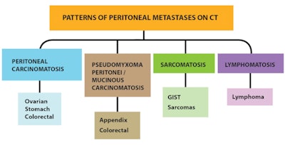
Depending on the primary tumor, different patterns of peritoneal seeding can be detected on CT. Also, abundant loculated ascites with scalloping of hepatic and splenic margins suggest pseudomyxoma peritonei (appendix, colorectal), while multiple solid masses of random distribution suggest sarcomatosis (gastrointestinal stromal tumor [GIST], sarcomas), they noted.
"Pseudomyxoma is a clinical concept that refers to abundant quantity of mucin inside the peritoneal cavity secondary to cancerous cells," the authors stated, adding that the most frequent origin is a low-grade appendiceal mucinous neoplasm.
The authors recommend bearing in mind the following clinical scenarios:
- A man in his fifth or sixth decade with a history of asbestos exposure and "dry" or "wet" carcinomatosis may have malignant mesothelioma.
- A woman in her fifth or sixth decade with a pattern similar to ovarian carcinomatosis but no adnexal mass may have primary peritoneal serous carcinoma.
- Patients with low-attenuation lymphadenopathy and previous history of granulomatous disease may have granulomatous peritonitis.
- A woman of reproductive age with obliteration of pouch of Douglas and hemorrhagic ovarian cysts may have endometriosis.

Radiologists must know about the pathogenesis of peritoneal metastases and its relationship with the peritoneal anatomy and the flow dynamics of the ascitic fluid, as well as the different patterns of presentation of peritoneal metastases on CT according to the type of primary tumor with surgical and pathological correlation, Miguez Gonzalez and colleagues recommended. Awareness of the differential diagnosis of peritoneal metastases compared with primary tumors of the peritoneum and nontumoral entities is important too.


