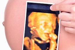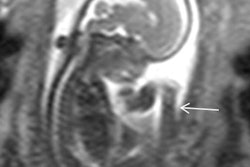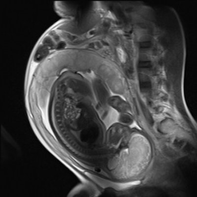
Use in utero MRI in conjunction with prenatal ultrasound scans when diagnosing fetal brain abnormalities -- that's the message from U.K. researchers, who found in utero MRI makes a "significant contribution" to the diagnosis by increasing accuracy when compared with ultrasound alone.
The group searched the literature for original studies on fetal MRI and prenatal ultrasound whose findings could be corroborated by postnatal imaging, surgery, or autopsy. The authors, led by Debbie Jarvis, senior radiographer at the academic unit of radiology at the University of Sheffield, found that using in utero MRI in tandem with ultrasound increased diagnostic accuracy by 16% than when ultrasound was used alone.
"When fetal brain abnormalities are suspected on ultrasound, in utero MR imaging is able to contribute significantly to the diagnostic pathway by both clarifying findings and increasing significantly the detection rate of abnormalities, particularly in midline and posterior fossa anomalies," they wrote (European Radiology, 21 September 2016).
Why MRI?
Fetal brain abnormalities occur in approximately 25 per 10,000 births in the U.K. and can result from environmental, chromosomal, genetic, or acquired causes, according to the authors. Accurate diagnosis of fetal brain abnormalities is necessary to guide management of the pregnancy and facilitate parental counseling, Jarvis and colleagues noted.
Ultrasound is the primary diagnostic imaging method for screening pregnancy and considered the reference standard for imaging the fetus brain, but there are occasions when technical limitations hinder clear visualization of the fetal anatomy. This led to investigating the utility of in utero MRI, but the true clinical value has not been established. Previous limited statistical evidence was unable to demonstrate any benefit of diagnostic accuracy -- until now.
The researchers sought to determine whether the diagnostic accuracy of in utero MR is superior, equivalent, or inferior to ultrasound by measuring the diagnostic accuracy of antenatal ultrasound alone (i.e., prior to in utero MRI) in relation to an outcome reference diagnosis determined by postnatal imaging, surgery, or postmortem examination, and measuring the diagnostic accuracy of in utero MR (following antenatal ultrasound) relative to an outcome reference diagnosis.
They included 34 studies in their analysis, totaling 959 cases, all of which had an outcome reference diagnosis determined by postnatal imaging, surgery, or autopsy. They found in utero MR imaging gave the correct diagnosis in 91%, which was an increase of 16% above that achieved by ultrasound alone (75%).
Ultrasound and MRI differed the most in regard to diagnosing midline abnormalities, particularly the posterior fossa, with MRI able to better detect abnormalities in the region.
However, in utero MRI is not without its limitations. Jarvis and colleagues also found the modality overestimates the presence of abnormalities more frequently than failing to identify them, which could be explained by the nature of fetal in utero MRI -- the need for fast imaging compromises image quality.
Also, to the untrained eye, artifacts from maternal breathing, fetal movement, and image aliasing may potentially mimic or obscure pathology.
"It is for this reason 'experience' should perhaps be defined by the number of fetal brain examinations reported," they wrote.
Timing also matters. The fetal brain develops rapidly and significant delay between ultrasound and MRI may influence the ability to diagnose accurately, because of natural brain development, increase in size of critical anatomical structures, or disease progression.
What's clear is more research needs to be done. The limitations of previous studies suggest further investigation is still required to clarify the full impact of in utero MR, the authors concluded.


