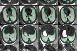
Fetal MRI is proving an increasingly useful technique that can provide valuable clinical information, and it should be carried out whenever a musculoskeletal (MSK) abnormality is suspected, according to Spanish researchers who have assessed over 1,000 pregnant women.
"Detection of musculosketetal fetal abnormalities is challenging, and false negatives are not rare," noted Dr. Amàlia González López, a radiologist at UDIAT-CD Hospital Parc Taulí in Sabadell. "The correlation between fetal MRI and clinical, radiological, and pathology studies is good and may help to understand MRI findings."
Fetal MSK abnormalities are common in syndromes or chromosomopathies with a poor prognosis, explained González López and her colleagues. They have used MRI to evaluate 1,025 pregnant women seen at their hospital between 1997 and 2017. Fetuses under investigation included those with MSK anomalies detected on MRI and could be correlated with clinical, surgical, or pathologic findings.
All exams were done on a 1.5-tesla system, with no sedation or special preparation, and the group presented their findings in an e-poster at RSNA 2017 that received a certificate of merit from the judges at the Chicago meeting.
MRI detected a total of 98 MSK abnormalities (see table). Of the 34 associated abnormalities, seven were of the central nervous system (CNS), five were abdominal, seven were thoracic, four were genitourinary, four were head and neck, two were of the spine, and five were in other regions of the body.
| Musculoskeletal abnormalities detected on MRI | |
| Limb and soft-tissue abormalities | No. of fetuses |
| Clubfoot | 54 |
| Hydrops | 13 |
| Reduction abnormalities | 11 |
| Arthrogryposis | 7 |
| Osteogenesis imperfecta | 2 |
| Polydactyly | 2 |
| Achondroplasia | 1 |
| Amniotic band syndrome | 1 |
| Arm dysplasia | 1 |
| Cervical mass | 1 |
| Ectridactyly | 1 |
| Genurecurvatum | 1 |
| Other positional abnormalities | 1 |
| Perineal mass | 1 |
| Thanatophoric dysplasia | 1 |
| Total | 98 |
These pregnancies resulted in a live birth in 58 cases, while interruption of pregnancy occurred in 24 cases and miscarriage was the outcome in five cases. MRI failed to detect 15 additional abnormalities, which were reported in the clinical, surgical, and pathology studies.
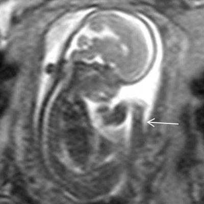
In clubfoot, the main risk factors are family history, intrauterine position, neuromuscular disorders, and oligohydramnios. Fetal MRI highlights malpositioning of the foot in thick slab T2-weighted sequences, explained the group. The second author of the RSNA e-poster, Dr. César Martín, is a senior pediatric radiologist who has worked on fetal MRI since 1997.
Genu recurvatum consists of congenital hyperextension of the knee associated with limited flexion, with or without dislocation of the knee, they continued. Depending on its cause, it can be divided into two types: malformative (anomalies of the elastic tissues) and postural (due to abnormal presentation or oligohydramnios). Associations for the malformative type are chromosomopathies and Larsen syndrome (flat face with hypertelorism, accessory carpal bones, and short terminal phalanges), and for the postural type are oligoamnios and footling presentation. On fetal MRI, positional abnormality of the knees can be seen in permanent unilateral or bilateral hyperextension, with the rest of the limbs in normal position.
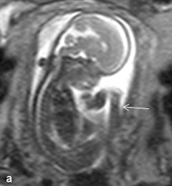
González López and her colleagues are convinced that MRI also has value in cases of thrombocytopenia-absent radius (TAR) syndrome, a rare condition in which thrombocytopenia is associated with bilateral agenesis of the radius. The associations consist of heart defects (33%, Tetralogy of Fallot and ventricular septal defects), facial anomalies (micrognathia, cleft palate), and partial bone defects in the limbs. Fetal MRI findings include absence of radius (with thumb present), radial deviation of the wrist (radial club hand), and variable shortening of the affected limb.
The technique can be useful too in amniotic band syndrome, which comprises a set of fetal malformations secondary to the entrapment of one or more fetal body parts by fibrous bands during gestation. The effects vary from slight indentations in soft tissues to amputation of limbs or fetal death. On fetal MRI, the bands are seen as linear tracts that are hypointense in T2-weighted images, reportedly identifiable in 57% of cases, and amputation and/or malpositioning of the limbs are visible, they stated.
In cases of popliteal pterygium syndrome, fetal MRI can find permanent flexion of the knee in relation to a membrane in the popliteal fossa, and cleft lip is one of the most common manifestations of this syndrome.
Arthrogryposis comprises a heterogeneous group of entities that give rise to multiple joint contractures, and restriction of fetal movement may be caused by neurogenic, myogenic, or extrafetal factors (amniotic bands or oligoamnios). Associations are talipes equinovarus, hip luxation, and CNS anomalies, such as agenesis of the corpus callosum, lissencephalia, and ventriculomegaly. Fetal MRI findings can include constant, abnormal position of a limb, and it most often affects distal segments of limbs, according to the authors.
Classifying limb deficiencies
Frantz and O'Rahilly were the first to categorize the various types of congenital skeletal limb deficiencies in the 1960s, and their classification is still of use today and includes the following:
- Terminal: the deformity extends to the distal region of the limb
- Intercalary: the distal region of the deformity is within the normal limits
- Transverse: involving both the radius and ulna or both the tibia and fibula
- Paraxial: involvement of only one bone in the limb
- Amelia: complete absence of a limb
- Meromelia: partial absence of a limb
- Hemimelia: partial absence of a large segment or half of a limb.
"Limbs (in hemimelia cases) are evaluated better in 3D T2-weighted HASTE [half-Fourier acquisition single-shot turbo spin-echo] sequences," González López noted.

