By using MRI to evaluate pregnant women with a short cervix on ultrasound, obstetricians can better predict if the expectant mother will give birth prematurely, according to a new study published online March 15 in Radiology.
Italian researchers used diffusion-weighted MRI and apparent diffusion coefficient (ADC) values to compare the inner subglandular zone and the outer stromal area of the cervix in pregnant women who were referred for suspected fetal or placental abnormality. ADC values from the subglandular zone were significantly greater in women who delivered their babies within seven days, compared with ADC values in mothers who delivered an average of 55 days later.
"In detail, subglandular ADC was inversely correlated to the interval between MR imaging and delivery; therefore, it emerged as a powerful imaging biomarker in the evaluation of patients with impending delivery," concluded lead author Dr. Gabriele Masselli and colleagues from Sapienza University of Rome.
Cervix measurements
Women in their second trimester of pregnancy with a cervix measuring 15 mm or less, as seen on ultrasound, are at greater risk of having a preterm birth. Ultrasound, however, is limited in its ability to predict a preterm birth because it does not provide information on changes in cervical tissue in the antepartum phase just before childbirth.
All 30 pregnant women in this study had a sonographically short cervix and a positive fetal fibronectin test between 23 and 28 weeks of gestation. Fetal fibronectin is a glue-like protein that helps hold the fetal sac to the uterine lining. Its presence before week 35 of gestation on a swab test may also indicate a higher risk of preterm birth.
Standard protocols
Two obstetricians at the hospital used ultrasound (Sonoline Elegra, Siemens Healthcare) and a 7.5-MHz vaginal probe to evaluate the uterine cervix. Three measurements were acquired of the cervical canal, with the average measurement used as the benchmark for each patient (Radiology, March 15, 2016).
The 30 pregnant women had a mean gestational age of 26 weeks and were found through their sonograms to have a short cervix of 15 mm or less.
MRI scans were conducted using a 1.5-tesla scanner (Avanto, Siemens) with a 16-channel phased-array torso coil covering the entire pelvis. The women were imaged in the supine position and entered the magnet feet first to minimize any feelings of claustrophobia.
The researchers calculated ADC values from MRI of the subglandular and stromal cervix -- subglandular ADC, stromal ADC, and ADC difference -- and correlated these parameters with the time interval until delivery. Receiver operator characteristics (ROC) curve analysis was performed to determine sensitivity and specificity of the ADC values in association with delivery within seven days.
ADC differentiators
Eight expectant mothers (27%) gave birth within seven days after admission to the hospital and their imaging scans (mean, 5 days; range, 3-7 days). They became part of the study's impending delivery group.
The other 22 women (73%) delivered their babies more than seven days after admission and scanning (mean, 55 days; range, 18-89 days) and became part of the late delivery group. Overall, 19 women (63%) had preterm delivery before 32 weeks of gestation.
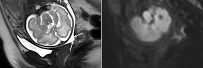 Images are from a 28-year-old woman at 30 weeks of gestation with a sonographic cervical length of 8 mm who was referred for suspected corpus callosum dysgenesis. MRI (left) shows a short cervix with funneling (arrows), with the cervical canal appearing as a zone of high signal intensity. Diffusion-weighted MRI (right) illustrates a uniform hypointense subglandular area of the cervix without any restriction of diffusion.
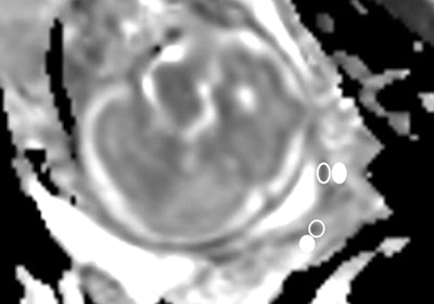 The ADC map gives a value of 2,681 x 10-6 mm2/sec in the subglandular area (empty oval). The patient gave birth four days later. Images courtesy of Radiology.
The researchers found no difference in cervical length on ultrasound between the two patient groups. ADC values in the outer cervical stroma were also similar between the groups.
However, the mean subglandular ADC and mean ADC difference were significantly greater in patients in the impending delivery group than in the late delivery group (p < 0.001). Subglandular ADC was found to inversely correlate with the time from imaging to delivery, the researchers noted.
"Our results indicate that a high ADC recorded at the level of the subglandular area of the cervix is associated with imminent delivery in asymptomatic patients who have a short cervix (≤ 15 mm)," they wrote.
Based on the ROC curve analysis, subglandular ADC had 100% sensitivity and specificity for impending delivery with a threshold of 1,921 x 10-6 mm2/sec.
Ongoing research
Masselli and colleagues noted several limitations of their study, including the small sample size. In addition, bias may have been introduced by limiting the study to patients referred for an MRI scan for suspected fetal or placental abnormality.
They plan to further investigate their results in larger, multicenter trials to confirm the role of subglandular ADC in predicting preterm birth.
"If this finding is confirmed in larger populations being studied for different risk factors, it could improve the policy of hospital admission and corticosteroid administration to improve fetal lung maturation," they speculated. "In addition, it could represent a new tool with which to monitor the efficacy of any drug putatively administered to slow the progression of impending labor."


