Measuring the minimal lumen area (MLA) of coronary arteries scanned at coronary CT angiography (CCTA) can help distinguish lesions that are hemodynamically significant from those that are not, reducing the number of false positives, according to a new study by researchers from Austria.
They found that a MLA cutoff of 1.9 mm2 or less showed the highest accuracy for predicting the hemodynamic relevance, potentially adding important value to CT angiography studies, the researchers reported at the 2014 RSNA meeting in Chicago.
 Dr. Fabian Plank from Innsbruck Medical University in Austria.
"We think this is a relevant parameter to indicate lesions that are highly suspicious for revascularization," said lead author Dr. Fabian Plank from Innsbruck Medical University in Austria.
Coronary CT angiography is a great test for ruling out coronary artery stenosis, but alone it cannot always distinguish flow-limiting coronary artery stenosis from those that merely look severe, according to Plank.
"What we've learned from over 15,000 patients in our internal registry is that CT angiography is an excellent gatekeeper to invasive angiography, and that patients without obstructive coronary artery disease had less than 8% risk of all cause mortality," he said. "On the contrary, patients with obstructive disease had an increased risk of all-cause mortality with the number of proximal segments, high-grade stenosis, and the number of positive segments or calcified plaques as the main predictors."
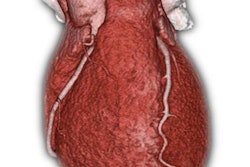
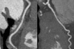
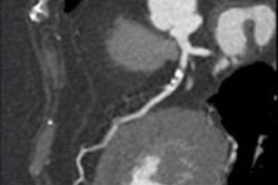
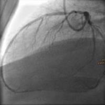
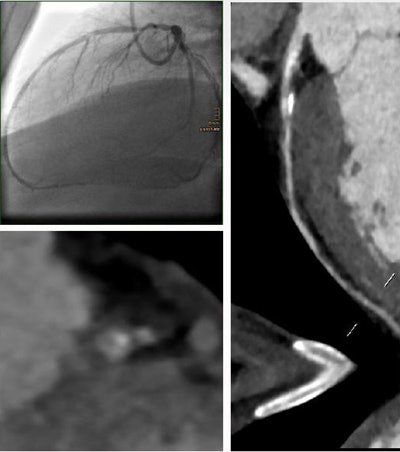 A 62-year-old smoker with hypertension, dyslipidemia, and diabetes had a 93% stenosis in the left anterior descending artery. The MLA was 1.4 mm2, the minimum lumen diameter was 1.1 mm, the area stenosis was 75%, and the diameter stenosis was 69%. The patient was revascularized. All images courtesy of Dr. Fabian Plank.
Visual grading has limitations
Clinical practice relies mainly on the visual grading of CT images to assess the severity of coronary stenosis, but there may be other, better approaches, such as quantitative measurement of stenosis, according to Plank.
"Therefore, we evaluated secondary measurements in CTA for the assessment of hemodynamic relevance and compared them to invasive angiography as our reference point," he said. The imaging was followed by coronary revascularization.
The retrospective study included all CCTA patients from the past nine years who had a high-grade stenosis at CCTA (≥ 50%) located in a proximal segment (right coronary artery, left main, left anterior descending, or circumflex artery) that had a minimum 5.5-mm lumen area.
A total of 80 patients (mean age 64.9 years) with 101 lesions in the proximal segments underwent either 128-detector-row (73 patients) or 64-detector-row CCTA (7 patients), followed by invasive coronary angiography. The MLA was quantified by CT along with minimum lumen diameter, maximal area stenosis, and maximal diameter stenosis.
All patients presented with at least one high-grade stenosis (> 50%) in a proximal coronary vessel, including the right coronary artery (RCA), left main (LM), left anterior descending (LAD), or circumflex (CX) artery, and subsequently underwent invasive angiography (ICA).
Results were evaluated for hemodynamic relevance in ICA (defined as fractional flow reserve less than 0.8 or stenosis 70% or greater) and followed by percutaneous intervention or coronary bypass grafting. Receiver operating characteristic (ROC) analysis with stepwise testing (0.1 mm2 MLA increments) also was performed.
According to the results, the optimal cutoff minimal lumen area was 1.8 mm2 or less to predict a positive invasive angiography result (above), This cutoff point yielded a sensitivity of 92.11% (78.6% to 98.3%), a specificity of 87.30% (76.5% to 94.4%), and an area under the curve (AUC) of 0.95 (0.91 to 0.99) for predicting stenosis significance at CCTA, Plank said.
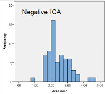 Results show that minimum lumen area is much larger for a lesion corresponding to a negative invasive angiography (above) versus the minimal lumen area for a positive invasive coronary angiography study (below). At the bottom, significance of results between positive and negative predictions.
"Similarly, the results of mininum lumen area, minimum lumen diameter, and stenosis were quantified, and we found a good cutoff value of 1.2 mm2 or a maximum stenosis of 60% when it comes to lumen diameter," he said.
Plank cautioned that obtaining a single diameter measurement "may not be enough for a reliable comparison" and that, surprisingly, the presence of calcification made no difference in the results or cutoff point.
As for study limitations, the interreader variability has not yet been calculated (two observers performed the measurement). Also, the reconstruction algorithm and image quality may affect the results -- for example. reduced image quality in CT of obese patients. Calcifications, too, may alter the results, and because the study included only proximal lesions, performance in distal stenosis is unknown, according to Plank.
"We found a very significant cutoff value of 1.8 mm2 minimal lumen area to predict relevant stenoses that are highly suspect to require coronary revascularization," he concluded.
But in addition to MLA, quantitative measurements should also include area and diameter of the stenosis and minimal lumen diameter, Plank noted.


