All healthcare staff have a responsibility to avoid unnecessarily high radiation dose, but the key players remain diagnostic radiologists who can and should integrate the necessary information and decide upon scan protocols, according to the lead author of an in-depth article published by European Journal of Radiology.
There are now many ways of cutting dose, including reducing tube current, tube potential, and improvements in image production, noted Dr. Takeshi Kubo, an assistant professor in the department of imaging and nuclear medicine at Kyoto University Hospital in Japan.
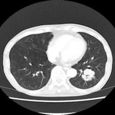
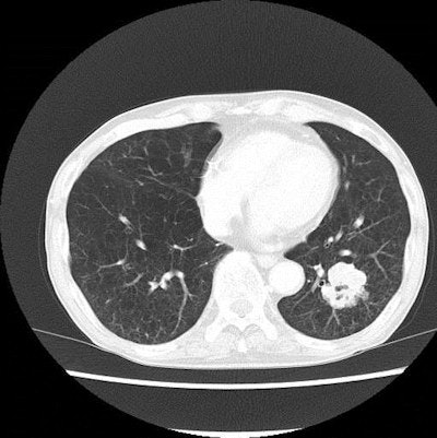
"We routinely use iterative reconstruction (IR) whenever possible, although the technique is not available on all scanners," he told AuntMinnieEurope.com. "Image quality is well-accepted now. There was some confusion at the texture change that IR caused, especially when comparison reading is requested, but now we are accustomed to the change."
 A pair of lung CT images of a patient with emphysema and lung cancer. Above, the image obtained with filtered back projection looks noisy. Below, the image produced with iterative reconstruction has improved noise profile while preserving the anatomical detail, which contributed to radiation dose reduction. Images courtesy of Dr. Takeshi Kubo.
The most notable change is the increased awareness of radiation dose among radiographers/technologists, who have been diligently working with radiologists to modify the scan parameter, Kubo added. Education is most important to secure patient safety, and without a sound understanding of the relationship between image quality and diagnostic yield, patients may fail to benefit fully from the advanced technologies.
"Radiologist shouldn't seek a 'pretty image,' but should try to understand what is sufficient for the particular patient," he advised.
Most medical radiation exposure comes from CT, and although CT scanners have improved technologically -- thus reducing CT dose -- there has been a boost in the number of exams, thus counterbalancing any benefits, wrote Kubo and colleagues in their comprehensive in-press article (EJR, 2014 July 9).
"There are varieties of measures that we can adopt to improve the safety of CT examinations," they explained. "We need to understand these methods and use them wisely to achieve dose reduction. Indeed, no single method works perfectly for all diagnostic needs. Users of diagnostic imaging should be aware of the advantages and disadvantages of these techniques so that they can use recent technical developments with minimal loss of diagnostic quality."
How can dose be reduced?
Radiation dose can be lowered by carefully following proper techniques such as correct patient positioning, appropriate scan length, and shielding the radiosensitive organs. For chest CT, shielding the mammary glands and the thyroid gland are applicable options. However, shielding could be harmful because of noise and artifacts, Kubo and colleagues noted. Scan parameters such as tube current and peak energy also can be adjusted for dose reduction. These too often come at a cost in image quality, though. Auxiliary measures that indirectly contribute to radiation dose reduction include alternative reconstruction algorithms or image filters. When the techniques are used with parameter modification to aid in radiation dose reduction, image quality is ameliorated.
Reduction of current-time product is the simplest method to reduce radiation dose to the patients, they noted.
"In principle, the radiation dose is proportional to the tube current if all other parameters are the same," they wrote. "Most often, tube current is modified to lower current-time product. Other factors, including gantry rotation time and helical pitch, also influence the current-time product."
High-pitch scanning lowers the radiation dose and on a single source scanner, pitch can be raised up to 1.5 without a gap in collected data. Dual-source scanners can tolerate a pitch of up to 3.6. The scan duration is shortened and, thus, so is dose. Plus, shorter scanning time is beneficial for patients who can't hold their breath for a long time.
"However, the image noise becomes higher with higher pitch if all other parameters stay the same," the authors noted. "Selection of pitch may alter other image characteristics such as z-axis resolution, artifacts. Therefore, as long as the duration of the scan is not critical issue, lowering tube current instead of raising pitch is a practical choice."
Many CT scanners are now able to automatically adjust the tube current according to body size as a part of automatic tube current modulation.
"Using constant tube current throughout the CT scan is not efficient because of the heterogeneity of attenuation values in parts of the human body," they stated. "If tube current is fixed, it needs to be set to the level where the worst image quality in the scan range still remains acceptable. Consequently, some parts may be excessively exposed to x-rays."
Automatic tube current modulation can work in 3D by combining two modes of current modulation -- current adjustment during gantry rotation control and current adjustment during table feed. The noise level of the image is primarily determined by the path of x-ray with the strongest attenuation where the highest tube current is needed.
Organ-based tube-current modulation can reduce the dose to radiosensitive organs close to the surface of the body, so when the tube comes closest to the mammary glands, for instance, the tube current is lowered.
There is no uniform standard of image quality, so the major vendors use their own method of image quality selection, such as reference mAs (Siemens Healthcare), standard deviation (Toshiba Medical Systems) noise index (GE Healthcare), and reference images (Philips Healthcare).
"It should be noted, however, that the dose-sparing effect would be dependent on the choice of the users," Kubo and colleagues wrote. "If you raise the target image quality the radiation dose goes up accordingly. Even with the use of automatic exposure control (AEC), we have to make sure that the image quality setting is appropriate to avoid unnecessarily high radiation exposure."
Another method to reduce CT dose is adaptive dose shield, which involves asymmetric control of the collimator, blocking nonproductive x-rays. The dose-saving effect varies with scan range: It's most remarkable in a scan performed with short scan range and high pitch, according to the authors.
Lower tube potential (kVp) also can reduce radiation dose. Switching to a lower kV will lead to significant reduction of photon fluence and, thus, a considerable increase in the image noise, they noted. However, radiation dose is proportional to peak kilovoltage to the power of 2.5. For instance, reducing kVp from 120 to 100 (17%) will lower the dose by 30%. When lower kVp is used, upward adjustment of the tube current is needed to preserve diagnostic accuracy.
"The unique advantage of low kVp use is the augmentation of the effect of iodine-containing contrast media, because average energy of x-ray becomes closer to K-absorption edge (33 keV) of the iodine," the authors wrote. "Actually, the low kVp technique is investigated mainly for contrast-enhanced CT, especially CT pulmonary angiography. ... It may be possible to attain radiation dose sparing with almost the same level of lesion conspicuity at the cost of increased dose of contrast material."
Iterative reconstruction also can lower radiation dose. Iterative reconstruction involves calculating projection data from reconstructed images, taking into account the effect of the scanner's physical properties. The calculated projection data are compared with measured projection data to correct projection data. The process is repeated to attain the final reconstruction images.
"With this method, higher CT image quality can be obtained with the same scan parameters," the authors noted. "Therefore, radiation dose reduction is possible without compromising the image quality."
Even with all the technological refinements, the choice of imaging procedures needs to be made through the "integration of clinical questions and sound knowledge of pathophysiology and imaging findings of the suspected condition," they stated.
Radiologists are still expected to play a role in the improvement of imaging practice to increase the efficiency in radiation use. Radiation dose reduction measures need to be repeatedly evaluated using objective image quality index and subjective assessment by the readers, but evaluation in terms accuracy and confidence level of the diagnostic performance are limited.
"More studies on the diagnostic accuracy and confidence level of radiation dose reduction techniques are needed, because the consistency of the diagnostic result is the most compelling evidence for the efficacy of imaging techniques," Kubo and colleagues wrote.
Radiation dose reduction in CT is a multifaceted issue, and understanding these aspects leads to an optimal solution for various indications of chest CT, they concluded.


