Austrian researchers added PET/CT to a package of three MRI sequences to create fused PET/MR images, which they found aided in the differentiation of suspicious breast lesions, according to a new study published online in Clinical Cancer Research.
Both FDG-based PET and MRI scans with multiple parameters have emerged as valuable tools in breast imaging, providing both functional and morphological information that improves sensitivity and specificity. Combining PET with multiparametric MRI could have widespread applications, but it has not been explored in great detail, according to a team led by Dr. Katja Pinker of the Medical University of Vienna (CCR, June 24, 2014).
 Dr. Katja Pinker from the Medical University of Vienna.
In what they called a feasibility study, the researchers decided to investigate whether fused PET/MRI scans, when used with three other MRI parameters, would improve the differentiation of malignant from benign breast cancer by enabling the quantitative assessment of multiple biological processes, including tumor angiogenesis, microstructural changes, increased glucose consumption, and cell membrane turnover. MRI parameters used were dynamic contrast-enhanced MRI (DCE-MRI), diffusion-weighted imaging (DWI-MRI), and 3D proton MR spectroscopy.
The group started with a patient population of 76 individuals, of whom 25 presented with clinical symptoms and 51 were asymptomatic but had screen-detected abnormalities. Patients underwent both multiparametric MRI scans on a 3-tesla scanner (Tim Trio, Siemens Healthcare) and PET/CT scans (Biograph 64 TruePoint, Siemens). PET and DCE-MRI images were fused semiautomatically on a workstation via software (TrueD, Siemens) using a landmark-matching tool, and color-coded images were generated.
The multiparametric MRI and PET/MRI data were evaluated by a breast radiologist and a nuclear medicine physician, with both readers assessing the probability of malignancy for each imaging parameter. Probability of malignancy was assessed using BI-RADS criteria. Reader accuracy with the three MRI parameters was then compared to accuracy with the three MRI parameters and PET/MRI.
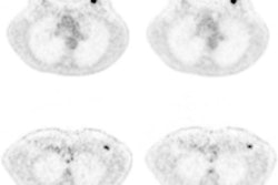
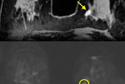
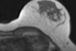
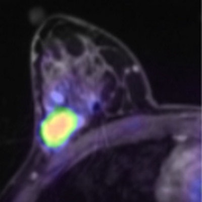
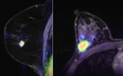 The left image shows invasive ductal carcinoma in a 48-year-old woman. Combined multiparametric F-18 FDG-PET/MRI accurately classifies the lesion as malignant. The irregular spiculated lesion demonstrates initial fast enhancement followed by a washout (BI-RADS 5), restricted diffusivity, and elevated choline levels, and it is highly FDG-avid (not pictured because only the fused PET/MRI image is shown). The right image shows invasive ductal carcinoma in a 39-year-old woman. The oval, circumscribed lesion demonstrates persistent enhancement (BI-RADS 3), restricted diffusivity, and elevated choline levels, and it is highly FDG-avid (again, not pictured). Three of four parameters were indicative of malignancy, and combined multiparametric F-18 FDG-PET/MRI accurately classifies the lesion as malignant. Images courtesy of Dr. Katja Pinker.
The four-scan protocol matching PET/MRI with multiparametric MRI had the highest sensitivity, at 100%, with specificity of 87%, resulting in a diagnostic accuracy of 96%, the highest among the techniques. Single-parameter DWI-MRI and 3D proton spectroscopy had a specificity of 96%, but there was a reduction in sensitivity ranging from 83% to 96%. Two-parameter imaging had higher sensitivity, but specificity ranged from 52% to 65%.
The four-scan technique also produced fewer biopsies, with 50% fewer biopsies compared to DCE-MRI alone and 38% fewer compared to DCE-MRI and another MRI parameter (although only the two-parameter comparison was statistically significant).
The authors concluded by stating that each of the tools used in the four-scan technique adds incremental value in diagnosing breast cancer, and combining all the information results in improved differentiation of benign and malignant tumors and reduced biopsies. They recommended that studies be performed with larger patient populations, as well as the development of a simple scheme for reading four-parameter PET/MRI studies.


