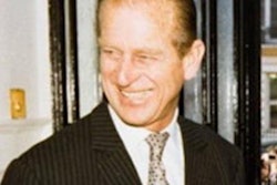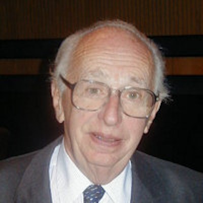
There has always been a close relationship between radiology and cardiology, and nothing shows this more than the life and work of Dr. Robert E. Steiner, who died peacefully on 12 September 2013.
Steiner worked at a time of dramatic changes in both radiology and cardiology, and he helped to train and inspire several generations of radiologists and cardiologists. It was in his department in the 1980s that I developed my lifelong interest in the history of radiology. The 1983 volume, Recent Advances in Radiology and Medical Imaging,1 was signed by those who recognized his outstanding achievements, and it marked his fourth decade in academic radiology.
 Dr. Robert E. Steiner (1918-2013).
Toward the end of a career devoted to cardiac and pulmonary radiology, Steiner developed an interest in MRI and made many contributions to this field. In recognition of his achievements, the MRI unit at Imperial College in London is named after him.2
Steiner was born in Prague in 1918, subsequently moving to Austria. He entered medical school at the University of Vienna in 1935 and in 1938 moved to Dublin, where he studied at University College, graduating in 1941. He retained affection for the Republic of Ireland throughout his life.
In the U.K., he worked for the Emergency Medical Services until 1945 and then started his radiology training at United Sheffield Hospitals. He was appointed to the staff of Hammersmith Hospital in 1950. He was made professor of diagnostic radiology at the University of London at the Post-Graduate Medical School, Hammersmith Hospital in 1960, a position he held until his retirement in 1983.
He was president of the British Institute of Radiology (BIR), the Royal College of Radiologists (RCR), and the Fleischner Society. He received many medals and awards, including the Gold Medal of the RCR, the Barclay Medal of the BIR, the Boris Rajewsky Medal of the European Association of Radiology (EAR), and the Gold Medal of the ECR and the EAR.
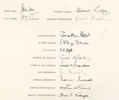 Signatures of professors of radiology who celebrated Steiner's contributions to academic radiology.
In 1972, his masterly presidential address to the BIR's annual meeting was on the impact of radiology on cardiology. This paper is still worth reading today.
He discussed the early days of radiology and celebrated the work of the Viennese pioneer Dr. Guido Holzknecht, who wrote the first textbook on chest radiology in 1901, by which time accurate interpretations of the cardiac contour could be made. Also, in 1929, Dr. Werner Forssmann published an account of the first cardiac catheterization in humans on himself and in 1933 the Swede Dr. Peter Rousthoi coined the term angiocardiography.
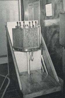
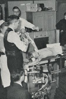 Left: Rotating drum serial changer, designed by Harry Coombs in 1948 in Sheffield. Cassettes are mounted on the drum, which can be rotated rapidly by hand between exposures. Right: Patient in front of rotating cassette drum for angiocardiography.
The next big breakthrough was in 1938 and 1939, when Drs. George Robb and Israel Steinberg reported the successful opacification of the of the right and left cardiac chambers and the pulmonary vasculature in acquired and congenital heart disease, stressing the possibility of using serial angiography coordinated with each heartbeat.
In 1941, Dr. André Cournand and colleagues showed that cardiac catheterization was a safe investigation in humans. Steiner's own interest in angiocardiography started in 1948, when together with Drs. John Goodwin and Sir Edward Wayne, he carried out the first angiocardiographic studies in Sheffield. In 1949, they were the first to demonstrate transposition of the great vessels in a living patient with congenital heart disease.
Steiner showed how advances in radiology profoundly influenced the practice of cardiology so that the cardiologist can correlate clinical findings with hemodynamic and anatomical information found at angiocardiography. He concluded this information has resulted in a more accurate interpretation of clinical findings. This was an exciting time in the history of radiology.
| Key dates in radiology and cardiology | |
| 1899 | Criegern: Indicates that fluoroscopy of the heart could provide valuable information. |
| 1899 | H. Reider and Jas Rosenthal in Germany outline the cardiac contour on a radiograph. |
| 1901 | Dr. Guido Holzknecht in Austria: The first dedicated book on chest radiology is published. |
| 1910 | O. Frank and W. Alwens in Germany injects a bismuth and oil suspension via the great veins into the heart of dogs and rabbits. |
| 1929 | Dr. Werner Forssmann in Germany performs first right heart catheterization by introducing a ureteric catheter into his own arm. |
| 1933 | Dr. Peter Rousthoi coins the term angiocardiography. |
| 1938/1939 | Drs. George Robb and Israel Steinberg report opacification of right and left cardiac chambers and pulmonary vasculature from a venous injection. |
| 1941 | Dr. André Cournand and colleagues show that cardiac catheterization can safely obtain haemodynamic information. |
| 1946 | Dr. Alejandro Celis in Mexico introduces selective intracardiac contrast injection. |
| 1951 | Drs. Elmo Ponsdomenech and Virgilio Beato Núñez perform left ventricular angiography using a direct needle puncture. |
| 1957 | Dr. J.B. Prioton in France performs left ventricular angiocardiography using a femoral artery approach. |
| 1958 | Dr. F. Mason Sones Jr. demonstrates that coronary vessels can be opacificed with contrast media. |
| 1977 | Dr. Andreas Grüntzig performs the first successful balloon dilatation of a coronary artery stenosis. |
In some respects, the 1970s can be seen as a golden decade for radiology with many new techniques becoming available. Steiner wrote in 1978 that who would have thought 10 years ago that such momentous developments were possible.4 There were many who felt diagnostic radiology had reached its peak and ultimate goal and there was no further room for new ideas and developments. How wrong they were in their ideas and predictions.
We are only at the beginning of the application of these newer techniques; it is quite impossible to predict how far they will advance our abilities to establish ever more accurate and definitive diagnoses. Without a doubt, the time will come when a reliable in vivo tissue diagnosis, possibly on a par with a histological diagnosis of biopsy, is possible. The questions then will have to be asked: How much further can we go? Should we advance our diagnostic refinements even further if they cannot be matched in the therapeutic field?
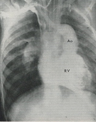 Venous angiocardiogram in a patient with transposition of the great vessels. The aorta (Ao) originates from the right ventricle (RV).
Unless advances in treatment go forward apace, further diagnostic performance will be of little help in patient management. Such thoughts at present are largely speculative and must not in any way influence our forward thinking in the advancement of the new imaging technology technologies and the application in our diagnostic endeavors.
The coming years augur well for diagnostic radiology, and there are exciting times ahead for all of us. How right Steiner proved to be.
Dr. Adrian Thomas is chairman of the International Society for the History of Radiology and honorary librarian at the British Institute of Radiology.
References
- Steiner RE, ed. Recent Advances in Radiology and Medical Imaging. Edinburgh, U.K.: Churchill Livingstone; 1983.
- Robert Steiner MR Unit. Imperial College London website. www1.imperial.ac.uk/medicine/divisions/cs/imagesci/facilities/hmr/. Accessed 16 September 2013.
- Steiner RE. The impact of radiology on cardiology. Br J Radiol. 1973;46(550):741-753.
- Steiner RE. Foreword. In: Kreel L, ed. Medical Imaging. Chicago, IL: Year Book Medical Publishers; 1979.
The comments and observations expressed herein do not necessarily reflect the opinions of AuntMinnieEurope.com, nor should they be construed as an endorsement or admonishment of any particular vendor, analyst, industry consultant, or consulting group.


