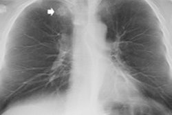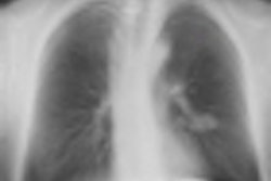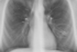
Using chest tomosynthesis with a digital radiography (DR) system reduced the number of chest CT exams performed at a Swedish institution, leading to lower radiation dose and better use of CT resources, according to a presentation from last week's RSNA meeting.
DR tomosynthesis creates 3D reconstructions of x-ray images acquired as the system's tube head pans around a region of interest. Tomo proponents believe the technology can be used to see around pathology that obstructs conventional DR images, allowing the use of lower-dose x-ray in some patients who previously were sent to CT.
For example, patients with lung nodules traditionally have been sent straight to CT after a suspicious chest x-ray. Now, the Swedish radiologists believe that some could be redirected to chest DR tomosynthesis.
The chest tomosynthesis unit at Sahlgrenska University Hospital in Gothenburg was introduced in 2006, and since then, more than 5,000 exams have been performed. The hospital has seen DR tomo's share of overall chest radiographs performed climb from 3% in 2007 to 7% in 2010.
As the use of chest tomosynthesis rose, the researchers began to wonder about the extent to which the technology could obviate the need for chest CT in some patients. In their RSNA presentation, they discussed a retrospective study that covered 100 radiologically and 100 clinically referred consecutive chest tomosynthesis exams.
Of the 100 radiologically referred exams, 86 were judged as beneficial for radiological workup, according to presenter Dr. Jenny Vikgren, from the department of thoracic radiology at the hospital.
"In 13 cases, the chest tomosynthesis adequately initiated further workup with chest CT," she said. Two radiologists judged the cases both independently and in consensus; they found that chest tomosynthesis alone dismissed the suspicion of tumors or nodules in 29 patients.
All of the 100 clinically referred chest tomosynthesis exams were judged as beneficial for the radiological workup. CT was avoided in 62 cases, she said. In 29 patients with nodules or tumors, tomosynthesis replaced CT as the follow-up modality, and in 15 patients, tomosynthesis was substituted for CT for at least one follow-up.
"In 11 patients, tomosynthesis was accepted as the primary staging modality for pulmonary metastases," she added. (The remaining seven patients had other diagnoses.)
Compared with chest x-ray, tomosynthesis added 0.12 mSv of radiation dose to each patient, but the avoided CT exams averaged about 4 mSv per patient.
When is the best time to use chest tomosynthesis?
"As a radiologist, when I look for the chest x-ray and I see a suspicion of tumor or nodule, I use chest tomosynthesis as a problem solver," Vikgren said.
If there is a nodule, it's discussed during a clinical conference and then the team decides whether to go ahead with CT, she added.


