MRI is proving significantly better than plain radiography for imaging of the medial extremity of the clavicle in forensic bone age estimation. In cases where accuracy is the primary concern, MRI should be used, but from a practical point of view, only in cases where radiography is inconclusive, at least for the time being.
That's the view of Elke Hillewig, a radiographer with a master's degree in criminal sciences who is currently performing PhD research at Ghent University in Belgium.
As more governments and nongovernmental organizations review their policies for age estimation of asylum seekers, increasing numbers of bone age examinations are being performed across Europe, but there are insufficient 3-tesla MRI machines for routine evaluation of the medial extremity of the clavicle.
"Controversy over the use of plain radiography still exists. Also, the use of ionizing radiation, as in CT and plain radiography in age estimation procedures, still remains problematic because of ethical and legal issues," Hillewig explained. "In the future, the use of MRI in forensic bone age estimation will probably increase. However, because of availability problems, it will probably be used as a supplement to plain radiography."
Several institutions are keen to start this procedure in the near future, she added. There is also increasing interest from sports medicine specialists looking to prevent fraud in junior competitions in which "nonjuniors" are included in "junior" teams.
At a seminar held in Brussels in December 2010, called "Unaccompanied minors: Children crossing the external borders of the EU in search of protection," Hillewig was moderator of an expert meeting about age estimation.
"There was certainly an international interest in new techniques for age estimation, such as our use of MRI for the evaluation of the medial extremity of the clavicle," she said. "It became clear that different organizations are looking to review their policies for age estimation of asylum seekers."
The meeting addressed the key principles on age assessment, the mutual recognition of age assessment, and the need for protocols and EU guidelines.
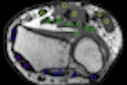
Hillewig's research is being promoted by Dr. Koenraad Verstraete, a professor and chairman of the department of radiology at Ghent University Hospital. The team in Ghent is creating a reference database consisting of 200 Caucasian males and females between the ages of 15 and 27. They're using conventional x-ray and 3-tesla MRI systems to study skeletal maturation versus age in the wrist and clavicle in these volunteers. The results are expected during the second half of 2011.
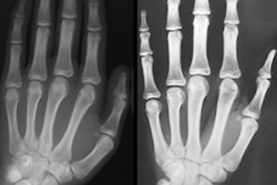
Her initial findings appear in the April 2011 edition of European Radiology. In the article, Hillewig explains according to recommendations of the international Study Group on Forensic Age Diagnostics (Arbeitsgemeinschaft für Forensische Altersdiagnostik, AGFAD), age estimations for living adolescents and young adults should consist of a physical examination (including anthropometric data, signs of sexual maturation, and potential age-relevant developmental disorders) and a radiograph of the left hand, as well as a dental examination, including recording of the dentition status and evaluation of an orthopantogram. At the age of 18, the hand ossification, third molar mineralization, and sexual maturation should be complete (Eur Radiol, April 2011, Vol. 21:4, pp. 757-767).
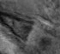
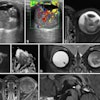
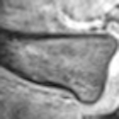
 High-resolution 3-tesla MR images of the medial part of the right clavicle. Stage I (left): There is no ossification center and the epiphysis is completely cartilaginous (high signal intensity). Stage II (right): There is a hypointense ossification center, which is connected to the diaphysis by hyperintense cartilage, without bony bridges.
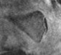
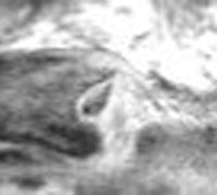
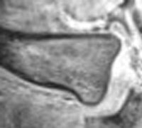 Stage III (left): In this early stage III, there is a hypointense ossification center, which is connected to the diaphysis by hyperintense cartilage, and a very small bony bridge, visible as a thin hypointense line between the epiphysis and the diaphysis. This small bony bridge is invisible on plain radiography. Stage V (right): Mature medial end of the clavicle. The ossification center has completely fused with the diaphysis, and the growth plate is invisible. All images courtesy of Elke Hillewig and Dr. Koenraad Verstraete.
Hillewig also recommends the work of Dr. Andreas Schmeling and colleagues at the Institute of Forensic Medicine in Münster, Germany, in this area. The Münster group published two cadaver studies on the subject that match Hillewig's results (Rechtsmedizin, December 2010, Vol. 20:6, pp. 483-488; International Journal of Legal Medicine, July 2007, Vol. 121:4, pp. 321-324).
