In an attempt to assist decision-makers in the migrant crisis, Turkish researchers have redoubled their efforts to identify an accurate way of evaluating bone age using pelvic radiographs in young adults.
"Most young victims of human trafficking and migrants in the European Migration Network are being documented as older than their accurate chronological age," noted Dr. Zehra Akkaya, from the department of radiology at Ankara University Faculty of Medicine. "To cope with many different legal and medical issues, a more accurate method for age determination for the Turkish population is required, especially after the influx of more than 1.5 million migrants, the majority of whom are younger than 18 years."
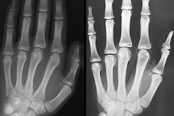
To determine age in young people, the most commonly used radiological studies are hand-wrist radiographs (including distal ulna and radial epiphysis), and elbow, shoulder, and pelvic radiographs that depict the iliac and ischial borders, explained Akkaya and colleagues in an e-poster at the 2016 congress of the European Society of Musculoskeletal Radiology (ESSR).
They designed a study to find dependable indicators to determine age using pelvic bony landmarks.
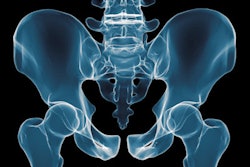
Anteroposterior digital pelvic radiographs of 698 individuals ages 18 to 22 and conducted between June 2011 and June 2015 were evaluated from the RIS/PACS at Ankara (RIS-i 5.0/Centricity PACS IW 3.7.3.9 SP3, GE Healthcare). A total of 277 (143 women and 134 men) were included in the study.
Radiographs lacking adequate diagnostic quality (e.g., due to inappropriate positioning or exposure), as well as radiographs of individuals with congenital or acquired pelvic or hip deformities, malignancies, surgical implants, prostheses, or signs of previous surgical interventions were excluded. Also, repeat radiographs of the same individuals were not included.
The hospital information system records were thoroughly evaluated for any known metabolic or systemic diseases that might affect bone development and maturation, and individuals with these conditions were also excluded.
The radiographs were evaluated simultaneously by two observers (one radiology specialist and one forth-year forensic medicine resident), and a consensus was reached about the degree of iliac crest and ischial tuberosity apophyseal ossification for both right and left sides on each radiograph.
For evaluation of iliac crest apophysis, well-established Risser signs consisting of six stages -- from stage 0 (no apparent ossification) to stage 5 (complete ossification and fusion of iliac crest apophysis) -- were considered and modified slightly because stage 4 was subdivided into groups 4a and 4b. Ischial tuberosity apophysis was evaluated for skeletal maturation too.
Because of the narrow age range (18-22 years), for the evaluation of ischial apophyseal maturation, the length of ossification at ischial tuberosity and its degree of fusion were assessed separately and subjects were divided into five groups as follows:
- Stage 0: No apparent ridge of ossified ischial apophysis
- Stage 1a: The ossified ridge was less than 50% of the obturator length
- Stage 1b: The ridge was more than 50% of the obturator length
- Stage 2a: Complete fusion of the ossified apophysis
- Stage 2b: Cortical irregularities in addition to stage 2a findings
The degree of fusion was determined according to the length of fusion between two arbitrary lines drawn perpendicular to the film at medial and lateral borders of the obturator foramen, which was referred to as the obturator length. The authors then compared the results with the chronological age and gender. No Risser stage 0-3 was encountered in this study and all individuals had a grade 4a or higher degree of iliac crest apophyseal maturation.
 Age and sex distribution of the study participants.
The table shows the age and gender distribution of the subjects. Chronological age was the most significant variable affecting the maturation degrees of iliac crests and ischial tuberosities, followed by gender (p < 0.05).
The maturation degrees of neither iliac crests nor ischial apophysis showed significant difference for right or left sides (p > 0.05), according to chronological age. But maturation degrees of both ischial tuberosities and right iliac crest showed significant correlation with respect to gender, with a positive (early maturation) for female gender (p < 0.05).
Both iliac crest and ischial apophyseal developments were significantly more advanced, especially for those ages 22 (p < 0.01). For the right iliac crest (p < 0.05) and both ischial tuberosities (p < 0.01), the developments were significantly more advanced in women compared with men.
Both iliac crest and ischial apophyseal developments were found to be reliable indicators to determine chronological age, but when iliac crest grading was compared with ischial grading, as individual variables, ischial tuberosity was statistically more accurate (p < 0.01), Akkaya and colleagues reported.
They found right iliac crest and both ischial apophysis to be reliable sites for the evaluation of bone age, most specifically to discriminate age 21 from age 22. The correlation of maturation degree of left iliac crest apophysis was not as accurate in age specification as the right side or either of the ischial tuberosities in the study.
"Although the retrospective design of the study limits us from determining the exact cause, one may propose that the dextrality may play a role in the earlier development on the right side," the authors stated. "Gender was found to be the second most important variable to affect bone maturation, with advanced degrees of maturation in females, especially for ischial tuberosities."
Overall, they found ischial tuberosities to be accurate sites to determine bone age, irrespective of the side (right or left) in the age range of 18-22 years old, as long as the gender of the individual was known beforehand. Also, if iliac crest apophysis is taken into consideration for identification of bone age, for this age range, the right side should be evaluated.
For more details and to view the authors' clinical cases presented at ESSR 2016, click here.
