Following the path of King Tut, Nefertiti, and other famed Egyptian figures, a collection of 500- to 1,000-year-old Chachapoyan mummies from northern Peru has now gone under the CT scanner -- a first in the study of this Andean people, according to research in the European Journal of Radiology.
Researchers in Austria used MDCT and 3D reconstruction to evaluate characteristics such as age, sex, stature, and disease in the group of 12 Chachapoyan mummies. Three objects from the burial site that were eventually identified as small animals were also imaged.
In addition to revealing physical characteristics, the study was intended to uncover details about "burial practices and other cultural aspects of this fascinating people," wrote Dr. Klaus Friedrich of the Medical University of Vienna and colleagues (Eur J Rad, available online August 6, 2009).
"Although several CT studies of Egyptian mummies followed by 3D reconstructions have already been described, until now no CT studies of Chachapoyan mummies have been performed," they wrote.
Peruvian past to present-day Vienna
The Chachapoyas, sometimes called the "people of the clouds," flourished in the cloud forests of the Amazonas region from the ninth century until the arrival of the Incas in the late-1400s, followed by Spanish conquest in the 1500s.
After being transported from the burial site in Laguna de los Cóndores to Vienna, the mummies underwent a single volumetric whole-body acquisition in spiral mode with a 64-slice MDCT scanner (Brilliance CT 64-channel, Philips Healthcare, Andover, MA). High-resolution scans of the temporal bones and teeth were also performed, and the three additional burial objects were scanned at standard resolution.
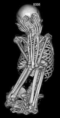 |
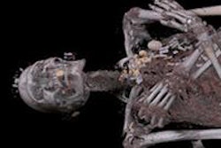
| A 13-year-old female mummy. Images courtesy of Dr. Klaus Friedrich. |
"First, the axial scans were evaluated, using different ranges of window width and window level parameters to identify structures with different densities," the authors wrote. Next, to virtually remove the bandages, "preset reconstruction algorithms for soft tissues with varying window width parameters were used to obtain the progressive elimination of external layers with a density lower than that of dried skin, starting from the bandages."
Anatomy and pathology
Sex and age of the mummies were determined by analyzing body structures such as the pelvis and skull, the authors wrote. Eight mummies were identified as female, three as male, and sex could not be determined for one. The ages of the mummies were newborn, 0.7 years, 2.5 years, 13 years, 13 years, and 16 years, with six mummies identified as being between 20 and 40 years old.
The height of the mummies, estimated by measuring femur and tibia length, ranged from 41 ± 3 cm (newborn) to 145 ± 14 cm (adult).
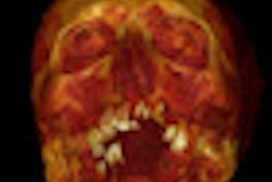
Dental conditions were compromised in seven of the mummies, including missing teeth, multiple carious lesions, and severe occlusal surface wear. The authors noted that damage was most likely due to a lack of oral hygiene, "but, especially chronic apical peridontitis, might in part also be explained by the excessive use of the coca plant (Erythroxylum coca) in the Chachapoyan culture, as in many other Andean populations, for religious and ritual purposes."
Among other findings, conditions identified in the bones included an occipital osteoma, as well as osteoarthritis or mild tuberculosis-related changes of a sacroiliac joint. A severe spondylodiscitis, a lytic lesion in the sacral bone, and erosions at one tarsal joint were also observed, which most likely represent severe manifestations of skeletal tuberculosis, the researchers noted.
Indications of pulmonary tuberculosis were also found. "Five of eight mummies showed several small intrapulmonary or mediastinal calcifications, such as those seen typically in pulmonary tuberculosis," the authors wrote.
Bundling and other burial practices
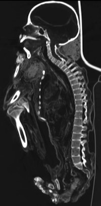 |
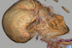
| This image, also of the 13-year-old female mummy, shows another view of the typical fetal position body placement. |
The researchers also surmised that the Chachapoyas specifically chose burial locations that protected the remains. Despite the rainy climate of the area, the ledge where the chullpas, or burial homes, were located resulted in a dry, cold microclimate that aided preservation.
The three additional burial objects were identified as skeletal parts of a mid-sized ape and two cats. "This on the one hand indicates the importance of those animals in the Chachapoyas' daily and ritual lives," while on the other hand underlining their ancestor worship, the authors wrote.
Friedrich and colleagues acknowledged the small number of mummies scanned and the fact that findings were not confirmed by other techniques as limitations of the study.
Regarding CT and anthropologic imaging, though, they noted that rapid improvements in CT technology have allowed for increasingly sophisticated multiplanar and 3D reconstruction options; these, in turn, have provided both improved quality and a greater quantity of information than could otherwise be obtained noninvasively -- a necessary approach for preserving the remains, they wrote.
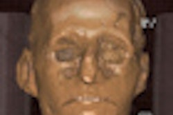
"Complex detector techniques and sophisticated postprocessing software led to an expansion of the paleoradiologic possibilities including virtual unwrapping of mummies, virtual fly through of different anatomic regions of the body, or reconstructions of the contours of the mummy's original face," according to the authors.
"However, most of the studies have been performed on Egyptian mummies; our study represents a unique MDCT dataset of Chachapoyan mummies," they concluded.
By Nicole Pettit
AuntMinnie.com staff editor
October 5, 2009
Related Reading
CT reveals Nefertiti's secrets, April 1, 2009
Philips CT images Egyptian mummy, February 5, 2009
Scan artist: Radiologist uses CT to reveal mystery of antiquities, October 25, 2005
CT helps unwrap mummy mystery, March 29, 2005
MDCT solves 400-year-old mystery, November 29, 2004
Copyright © 2009 AuntMinnie.com


