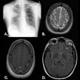
NEW YORK (Reuters Health), Jul 24 - Whole-body MRI appears useful in assessing acute inflammatory lesions in the sacroiliac joints in patients with established and active spondylarthritis (SpA), European and U.S. researchers report in the July 15 issue of Arthritis and Rheumatism.
The approach, lead investigator Dr. Ulrich Weber told Reuters Health, "is a promising tool to assess the inflammation load in the entire axial skeleton of SpA patients -- as opposed to regional exams by conventional MRI."
Weber of Balgrist University Hospital, Zurich, Switzerland, and colleagues came to this conclusion after using both methods in 32 patients. The MRIs were then scored independently in random order by three blinded readers.
"The performance of whole-body MRI in assessing sacroiliac joint inflammation," continued Weber, "compares well to conventional regional MRI of the sacroiliac joint."
In the same issue of the journal, Weber along with Swiss and Canadian colleagues also report on whole-body MRI in assessing spinal inflammatory lesions in patients with ankylosing spondylitis or with inflammatory back pain. He noted that "the same equivalence has been shown for spinal inflammatory lesions."
The team used the same technique to evaluate 35 patients with ankylosing spondylitis, 25 with recent-onset back pain, and 35 healthy controls. All were 45 years old or younger.
Again, the images were interpreted by three blinded readers. The investigators found that diagnostic utility was optimal when two or more vertebral corner inflammatory lesions were recorded.
For ankylosing spondylitis patients, the approach provided a sensitivity of 69%, a specificity of 94%, and positive likelihood ratio of 12. In the back pain patients, the corresponding values were 32%, 96%, and eight. Nine of the controls had one or more vertebral corner inflammatory lesions, but only two had more than two.
"An MRI finding of two or more corner inflammatory lesions in the spine in the age group below 45 years makes a diagnosis of SpA highly probable," Weber said. "Single corner lesions can be misleading, as one quarter of healthy individuals showed such a signal alteration."
By David Douglas
Arthritis Rheum 2009;61:893-908.
Last Updated: 2009-07-23 18:57:58 -0400 (Reuters Health)
Related Reading
MRI sees spinal metastases earlier, but low-dose whole-body MDCT aces skeletal workup, May 20, 2005
Copyright © 2009 Reuters Limited. All rights reserved. Republication or redistribution of Reuters content, including by framing or similar means, is expressly prohibited without the prior written consent of Reuters. Reuters shall not be liable for any errors or delays in the content, or for any actions taken in reliance thereon. Reuters and the Reuters sphere logo are registered trademarks and trademarks of the Reuters group of companies around the world.
