Venous disease is widespread and common, and it is particularly topical right now, given the potential side effects of COVID-19 vaccines. In a Q&A interview, Prof. Thomas Vogl outlines which venous disorders his team deals with in their daily clinical routine and how they diagnose and treat patients.
Vogl is head of the Institute for Diagnostic and Interventional Radiology at the University Hospital Frankfurt am Main and president of the 102nd German Radiology Congress, which takes place from 27 March to 8 November 2021. The interview was conducted by the German Röntgen Society (Deutsche Röntgengesellschaft, DRG).
Q: Which venous disorders do you deal with most frequently in your daily medical practice?
A: The human venous system and the diseases that can manifest themselves are an important field of activity for interventional radiology. Interventional radiology offers patients not only diagnostics but also minimally invasive therapeutic interventions using imaging.
 Prof Dr. Thomas Vogl.
I think a lot of people don't even know what interventional radiology can do in this area. At our institute, we very often have to deal with patients who suffer from thrombosis. These blood clot occlusions most commonly occur in the veins in the leg. In diagnostics, we clarify thromboses and possibly resulting pulmonary embolisms. The topic of thrombosis is currently very much publicized by reports on the AstraZeneca COVID-19 vaccine and sinus vein thrombosis.
Patients also come to us who suffer from varices, i.e., superficial vein enlargements that are "knotty" and clearly visible through the subcutaneous tissue. Such varices or varicose veins show up in women and men. In the case of internal vein problems, for example, we see enlarged veins that can press on nerves in the pelvic area of affected women. This condition can also occur in men, with the result that their fertility can be severely impaired. What also plays a very important role in my everyday radiology practice and in my institute are venous diseases as a result of venous malformations, which we have to evaluate.
Q: Which imaging methods do you use at your institute to diagnose venous disease?
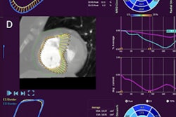
A: The first method is ultrasound, which can determine the velocities in the venous system very precisely. The course of the vein is well documented, and it also allows the detection of superficial and medium-depth diseases of the veins. If pathologies become apparent, we use advanced imaging methods, particularly CT. This rules out a pulmonary embolism, documents the course of the vein, and determines the extent of thrombosis. With MRI, sinus vein thromboses can be diagnosed very well.
Q: On which venous disorders do you work therapeutically as an interventional radiologist?
A: In principle, interventions on the veins do not differ significantly from interventions in other diseases. Interventional radiologists treat chronically ill patients, for example by inserting port systems into the veins. In addition, when it comes to veins, we also deal with the aforementioned venous malformations and thromboses.
 Catheter in subclavian vein. Left subclavian with tumor stenosis. Image shows large-lumen 9-mm balloon after expansion of the stenosis and before the stent is applied. Courtesy of Prof. Dr. Thomas Vogl and University Hospital Frankfurt.
A typical venous therapy is, for example, one we use on young men who have impaired fertility. The cause of this condition is dilated veins in the testicles and kidneys. Interventionally, we do it in such a way that we embolize these vein clusters and thereby restore the fertility of these -- quite often -- young men. We also remove foreign bodies from veins. To do this, we use instruments such as pliers, wires, and drum/barrel devices.
Q: What interventional methods are used for thrombosis?
A: In the case of thromboses, we offer the option of removing thrombi in interventional radiology. We reopen vessels that have become diseased due to chronic occlusion caused by thrombosis, and we can insert stents there to achieve outflow, for instance. We access mainly via the blood vessels -- i.e., arteries or veins -- with the help of catheter technology.
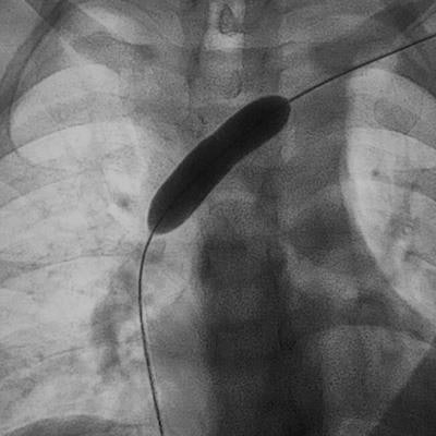
 60-year-old male patient with severe stenosis after stent angioplasty of the right brachiocephalic vein. Images courtesy of Prof. Dr. Thomas Vogl and the DRG.
Q: Does interventional radiology also play a role in patient follow-up?
A: Chronic diseases of the veins in particular have to be checked again and again through imaging. This is especially true for venous malformations or when stents are placed in the veins. Unfortunately, diseases of the veins are usually chronic. If not handled carefully, they can lead to physical disfigurements such as varices. Above all, venous disease can have very dangerous consequences for the cardiovascular system. That is why the early detection of thromboses, tumors, or venous malformations is essential.
Editor's note: This is an edited translation of an article published in German by the DRG on 22 April 2021. Translation by Frances Rylands-Monk. To read the original version, go to the DRG website.