Male professional golfers have an average age of only 24, and along with their different swing mechanics and increased repetitive strain, this factor results in injury patterns that vary considerably from those of the amateur, according to research presented at the 2015 congress of the European Society of Musculoskeletal Radiology (ESSR).
"A working knowledge of the complex swing mechanics, along with precise history-taking and deft examination, is essential in formulating a diagnosis and requesting the correct radiological test," noted Dr. Amit Kumar Bharath, a radiology registrar at the University of Leeds University Hospital in the U.K.
Golf remains a popular sport, and is growing particularly fast in China, where around 20 million players are expected by 2020. However, the frequency and nature of wrist injuries in elite golfers has not been reliably documented until recently, Bharath and colleagues explained.
 A pro golfer's swing imposes great strain on the wrist.
The top pro golfers have reported that most injuries are sustained in the leading wrist, with ulnar-sided injuries being commonly reported. Complex swing mechanics are thought to subject the leading wrist to considerable stress as it moves from ulnar to radial deviation and then back to ulnar deviation during the swing (see box below for more details). There is also movement from pronation to supination, and from flexion to extension through impact.
"This impact is substantial as professionals aim to 'hit through the ball' with the club face, often hitting the ground and taking a divot of turf in order to make more controlled shots through imparted spin," they wrote.
Wrist injuries under scrutiny
The authors studied the common wrist injuries encountered by the examining sports physician in the elite golfer. Injuries to the wrist can be classified by the anatomical localization of pain and other symptoms, and common ulnar-sided, radial-sided, and dorsal injuries must also be evaluated, they pointed out.
Up to 67% of wrist injuries occur in the ulnar aspect of the lead wrist, which is the left wrist in a right-handed golfer. The extensor carpi ulnaris tendon is involved in the majority of ulnar-sided injuries, and typical tendon pathology includes subluxation, dislocation, tendonopathy, and tenosynovitis. Dynamic ultrasound during pronation and supination can evaluate the position of the extensor carpi ulnaris tendon within the ulnar groove.
The true incidence of stress-related fractures in golfers is unknown, but a fracture of the hook of the hamate is a common ulnar-sided wrist injury, and after rib fractures, it is the most common stress fracture seen in golfers, according to Bharath et al. CT is the best modality for evaluating occult hamate fractures, with a reported sensitivity of 100% and a specificity of 94% compared with 72% and 88% in plain radiographs. Associated injuries of hook of hamate fractures include injury to the ulnar nerve and artery, and damage to the flexor carpi ulnaris tendon, they added.
Dorsal rim impaction syndrome, also known as hypertrophic synovitis, is a type of carpal impingement syndrome that predominantly affects the trailing wrist in elite golfers, and dorsal impaction syndromes are also seen in athletes, notably gymnasts, weightlifters, and those who do excessive press ups, the authors continued. Plain x-ray and CT are usually normal as this condition, at least initially, is a capsular soft-tissue abnormality. Dorsal capsular thickening can be shown on ultrasound or MRI, but MRI is more reliable in documenting its extent as well as any concomitant injuries or bone marrow changes elsewhere in the wrist.
"A carpal boss is the description for a bony protuberance localized to the carpometacarpal region at the base of the index and middle finger metacarpal bones (the quadrangular joint)," they pointed out. "Bossing can result in significant morbidity and time away from practice and play for the golfer."
Plain x-ray can be diagnostic for its presence, but if negative because of bony overlap, CT or MRI can define the bossing. MRI should be performed routinely in elite golfers as this technique can also demonstrate associated bone marrow edema, fracture, soft-tissue edema, and/or tendon abnormality, the Leeds group recommended.
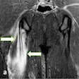
Finally, dorsal ganglion cysts of the wrist are the most common focal lesions in the hand and wrist in the general adult population, and are also common in elite golfers. On MRI, ganglia are typically fluid signal being low signal on T1-weighted images and high signal on T2-weighted images, but a high proteinaceous content or hemorrhage can result in lesions appearing isointense or hyperintense on T1-weighted images.
A narrow stalk connecting the ganglion to the joint is often visualized. The sonographic findings mirror MRI, and typically a hypoechoic fluid-filled mass is seen with a narrow stalk extending toward the joint. An elongated neck can cause the ganglion to surface at distance from the joint and appear at an apparent atypical location, they concluded.
The pressure points of the golf swing
The wrists link the body to the golf club and form the final component of a kinematic chain composed of the hips, spine, and shoulders. An overview of wrist movement through the swing does however aid our appreciation of the stresses incurred at the wrist and provides a useful framework on which to correlate pathology.
The nondominant wrist (left wrist for right-handed golfers) begins the golf swing in a position of ulnar deviation when addressing the ball. As the club is lifted away into the backswing, this wrist moves into radial deviation until it sits maximally radially deviated at the top of the backswing. At this point, the club changes direction to begin the downswing and the nondominant wrist returns to ulnar deviation until the ball is hit.
The dominant wrist moves in a different path altogether, being in neutral at address before moving quickly into maximal extension during the backswing and only coming back into neutral (and ultimately flexion) just before ball strike. In this way, the wrists control the golf club and impart significant power to the ball strike. The limited joint movement ensures consistency and increased reproducibility in the swing.
These opposite motion paths (ulnar/radial deviation for the nondominant wrist, flexion/extension for the dominant wrist) allow an understanding of the incidence of certain injury patterns in golf. The ulnar and radial deviation movements of the leading wrist are likely to predispose to problems with the tendons on the radial and ulnar borders of the wrist as they move through a near-maximal excursion with these wrist movements. This explains the high incidence of de Quervain's tendonitis and extensor carpi ulnaris sheath and tendon problems in the nondominant hand.
(Source: Dr. Amit Kumar Bharath, presented at ESSR 2015).


